Case
A pregnant woman aged 31 years who was 6 weeks pregnant presented with erythematous, scaly, annular plaques in her pubic region that spread to her torso and limbs 4 weeks after a holiday to India (Figure 1). This was initially diagnosed as eczema, and the patient was commenced on betamethasone diproprionate 0.05% and hydrocortisone 1% creams. Lack of response prompted the clinical suspicion of tinea corporis, and she received 3 weeks of treatment with terbinafine 1% cream followed by 2 months of clotrimazole 1% cream, without clinical improvement. A skin scraping was collected for fungal microscopy and culture. Trichophyton indotineae was cultured.
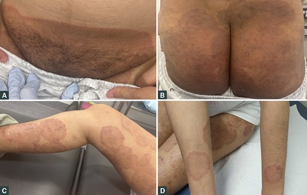
Figure 1. Extensive tinea corporis with erythematous, scaly, annular plaques on initial presentation following treatment with topical terbinafine and topical clotrimazole creams. A. Pubic region. B. Buttocks. C. Legs. D. Arms.
Question 1
What is the clinical significance of T. indotineae?
Question 2
How is tinea corporis treated and how is treatment different for T. indotineae?
Question 3
How do you diagnose tinea and what diagnostic investigations would be beneficial for treatment-resistant tinea infections?
Question 4
What are the considerations of treatment given the patient’s pregnancy and future plans to breastfeed?
Answer 1
T. indotineae is a newly recognised species of dermatophyte that demonstrates high terbinafine resistance. Normally, terbinafine exerts a fungicidal effect by inhibiting squalene epoxidase, causing ergosterol depletion and toxic squalene accumulation leading to the disruption of fungal cell membranes and then cell death.
1
The global spread of T. indotineae has followed initial outbreaks in the Indian subcontinent. Widespread use of over-the-counter high-potency steroid and steroid–antifungal combination creams has led to perceived infection suppression, which, along with incomplete or inappropriate treatment with readily available systemic anti-fungal agents, has fostered chronic infections and selected for resistant T. indotineae.2 It presents as a generalised inflammatory and pruritic tinea corporis. Australian cases remain limited, with T. indotineae accounting for 6.4% of non-nail specimens submitted for dermatophyte culture to a Melbourne laboratory.3
Answer 2
First-line treatments for tinea in Australia are topical or oral terbinafine.4 Systemic therapy is recommended for those with hair follicle involvement, nail involvement, failure of topical therapy, extensive disease or recurrence. As T. indotineae is highly resistant to terbinafine, itraconazole is recommended for suspected or proven infections.5 Itraconazole is highly lipophilic, leading to variable bioavailability across formulations. The super bioavailable (SUBA) itraconazole (Lozanoc, Mayne Pharma International Pty Ltd) is preferred because of its higher and more consistent absorption. The recommended adult dose is SUBA itraconazole 100 mg twice daily (bd). If unavailable, conventional itraconazole 200 mg bd (eg Itracap, Alphapharm Pty Ltd trading as Viatris; Itranox, Arrotex Pharmaceuticals Pty Ltd) can be used. Because of its long half-life, steady state is reached slowly, and trough (pre-dose) concentration level should be checked after 10–14 days, aiming for a therapeutic range of 1–4 mg/L to ensure efficacy and minimise toxicity.6 A duration of 2–12 weeks based on clinical response is recommended;5 however, we recommend continuation of therapy with appropriate monitoring until the patient is clinically and mycologically clear from tinea infection.
Answer 3
Tinea corporis typically presents as single or multiple rapidly spreading, asymmetric annular erythematous plaques with a raised scaly edge. When multiple plaques are present, the distribution of tinea corporis might be bilateral, but it is not typically symmetrical. In lightly pigmented skin, lesions tend to be erythematous, whereas in skin of colour, lesions might appear either hypopigmented or, when chronic, hyperpigmented (Figure 2B and 2C). Untreated chronic tinea corporis can also lead to confluence of lesions and a psoriasiform or eczematous appearance in skin of colour (Figure 2D). The differential diagnoses of tinea corporis are presented in Table 1. If the diagnosis of tinea is in question, a skin scraping for fungal testing should be taken before any treatment is commenced.7 If tinea is suspected clinically on skin examination, it is essential to examine the scalp and nails (and take a hair pluck and nail clip, respectively), as infection at either of these sites will upgrade treatment from topical to systemic anti-fungal therapy.
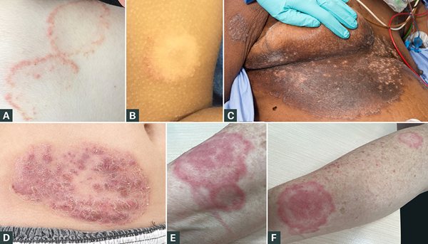
Figure 2. A. Tinea corporis on the back of a patient with lightly pigmented skin. B. Hypopigmented tinea on the upper arm in a patient with skin of colour. C. Chronic hyperpigmented tinea on a patient with skin of colour, with confluence of lesions creating a psoriasiform appearance. D. Tinea incognito on the abdomen presenting as Majocchi granuloma reflecting deeper infection with hair follicle involvement. E., F. Tinea incognito on the right forearm presenting as tinea pseudoimbricata with multiple concentric lines.
| Table 1. Comparing common differential diagnoses for tinea infections on the basis of their morphological features, distribution and associated findings |
| Condition |
Morphological features |
Distribution |
Associated findings |
| Discoid eczema |
Pruritic annular erythematous plaques – often exudative/crusted in the acute phase and scaly and lichenified in the chronic phase |
Typically bilateral and symmetrical |
Often personal and/or family history of atopy |
| PsoriasisA |
Well-demarcated plaques with silvery- white scale. Plaques are erythematous in lightly pigmented skin but often more violaceous to hyperpigmented in skin of colour |
Typically affects extensor surfaces and scalp but might demonstrate an inverse pattern |
- Can have nail and/or joint involvement
- Family history of psoriasis is often present
|
| Pityriasis rosea |
Oval, salmon-coloured papules or plaques with collarette scale. Initially ‘herald patch’ prior to generalised eruption |
Mainly affecting the trunk and proximal limbs; ‘Christmas tree’ pattern following Langer’s lines |
- Often results in hypo/hyperpigmentation that takes months to resolve
- Might follow a viral infection or can be drug induced
|
| Granuloma annulare |
Smooth, skin-coloured to pink papules forming annular rings with central clearing, generally non-scaly |
Can be localised or generalised depending on subtype; however, it commonly affects dorsal hands, feet and elbows |
Can be associated with autoimmune diseases (ie thyroiditis, diabetes mellitus) and hyperlipidaemia |
| Contact dermatitis |
- Erythematous, oedematous papules, vesicles or plaques
- Might become crusted or lichenified with chronicity
- Allergic contact dermatitis is intensely itchy
|
- Site of maximal involvement corresponds with area of contact with the irritant/allergen
- Allergic contact dermatitis might display autoeczematisation (Id reaction, autoreactivity, generalisation) to other areas
|
- Thorough history should be able to identify exposure to irritant/allergen
- Allergen avoidance prevents recurrence
|
| Fixed drug eruption |
Well-defined solitary or multiple round/ oval erythematous to violaceous plaques, sometimes with bulla formation |
- Most frequent on the hands, feet, eyelids and anogenital and oral mucosa
- Can uncommonly be generalised
|
- Lesions heal with post-inflammatory hyperpigmentation
- Thorough history should be able to identify culprit drug exposure
- Re-exposure leads to recurrence – typically with shorter lag-time and more severe presentation
|
| ImpetigoB |
Can present as erythematous papules/ pustules that rapidly crust over (crusted impetigo) or vesicles/bullae with turbid fluid that rapidly erode (bullous impetigo) |
Mostly found on the face and on other sites of exposed skin |
- Highly contagious and often cluster in school and home environments
- Can occur secondary to pre-existing skin conditions (ie eczema)
|
| FolliculitisA |
- Perifollicular erythematous papules/ pustules, sometimes coalescing
- Pruritic and/or tender
|
Affects hair-bearing sites, commonly the scalp, beard, trunk, buttocks and thighs |
Commonly associated with infections (bacterial, fungal, viral), occlusion and irritation |
A These conditions are more common mimics of tinea incognito where steroid use has altered their morphology.
B This morphology is commonly seen in zoophilic species of tinea infections. |
The inappropriate use of topical steroids can lead to tinea incognito, where the classic features are altered, including reduced scaling, loss of or exaggerated annular morphology, papule or pustule formation (reflecting hair follicle extension of infection, known as Majocchi granuloma) and multiple concentric lines (known as tinea pseudo imbricata; Figure 2E and 2F). The use of potent topical steroids can also cause skin atrophy, telangiectasia and striae, and application to the skin prior to fungal testing significantly reduces the yield of fungal scrapings, resulting in false-negative results.
Culture-based diagnosis remains the most common method used, providing the benefit of an isolate for susceptibility testing.4 Rapid turnaround time can be achieved with polymerase chain reaction (PCR)-based testing;8 however, in Australia, the most widely used assay (Dermatophytes and other Fungi, AusDiagnostics) cannot currently differentiate T. indotineae from other members of the T. mentagrophytes/T. interdigitale species complex.4 This importantly highlights the need for clinician education and recognition of those who might be at risk of T. indotineae to ensure that specific testing for T. indotineae is requested, especially in cases of returned travellers from endemic areas such as India and those with treatment-resistant infections. This case had both features (a returned traveller from India and treatment-resistant disease), with the diagnosis confirmed following dermatophyte testing. As an emerging problem and not yet endemic, it highlights the importance of a travel history in helping to guide suspicion of a diagnosis of T. indotineae. Both culture and PCR remain important in fungal diagnostics for cases where T. indotineae is suspected and in treatment of refractory cases. In these cases, further testing for specific identification should be undertaken, with consideration of whole-genome sequencing to identify genotypic correlates of resistance.
Answer 4
Oral antifungal treatments are not recommended during pregnancy and are preferably delayed until postpartum, with avoidance in the first trimester as a minimum.9 Itraconazole safety data are limited; however, foetal outcomes after maternal exposure to oral antifungal agents have shown there might be an increased risk of eye defects, which should be monitored carefully.10 Oral antifungals are not recommended during lactation because of their often-long treatment time and should be avoided, if possible.11 When indicated, consultation with specialists such as infectious diseases, dermatology and obstetric physicians is recommended to ensure the safety of both the patient and their developing foetus.
Case continued
After being referred to a tertiary dermatology service and following consultation with infectious diseases and the patient’s obstetric physician, she was commenced on itraconazole 100 mg po bd, short-term combination hydrocortisone 1%/ clotrimazole 1% cream for pruritus, and ketaconazole 2% shampoo as a shampoo- and-wash to minimise contagion. The itraconazole dose was optimised to 200 mg bd, guided by her serum levels. Following 22 weeks of treatment, clinical improvement in itch and scaling was observed, with post-inflammatory hyperpigmentation persisting (Figure 3). It is worth noting that post-inflammatory hyperpigmentation is common in skin of colour, arising when inflammation stimulates excess melanin production or dermal pigment trapping. It is recognised by smooth, macular hyperpigmentation in areas of prior disease activity.
Repeat PCR testing from skin scrapings was negative for T. indotineae, and treatment was ceased. The extended treatment duration of 22 weeks was attributed to relative immunosuppression caused by pregnancy,12 as well as initial cautious dosing in the context of the altered volume of distribution and concerns of potential maternal–foetal toxicity.
Clinicians must be aware of the increasing emergence of T. indotineae, especially in returned travellers. Where clinically suspected, further testing for specific identification is warranted. Because of the limitations in laboratory testing in Australia, T. indotineae might be underdiagnosed, and prompt recognition and treatment will mitigate the risk of further spread of this dermatophyte in Australia.
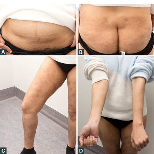
Figure 3. Clinical improvement of the patient following 22 weeks of itraconazole treatment with areas of post-inflammatory hyperpigmentation. A. Pubic region. B. Buttocks. C. Legs. D. Arms.
Key points
- T. indotineae is a highly resistant strain of dermatophyte infection that has spread to Australia, causing an extensive disease that is refractory to the common first-line treatment of terbinafine.
- Given its high degree of resistance to terbinafine, the use of itraconazole is recommended as first-line treatment in patients with high clinical suspicion of or confirmed indotineae infection.
- Clinicians should be aware of this emerging dermatophyte in Australia, and where there is clinical suspicion (ie returned traveller, extensive inflammatory tinea, treatment resistance), skin scrapings for fungal microscopy and culture (±PCR) should be collected for analysis.