Case
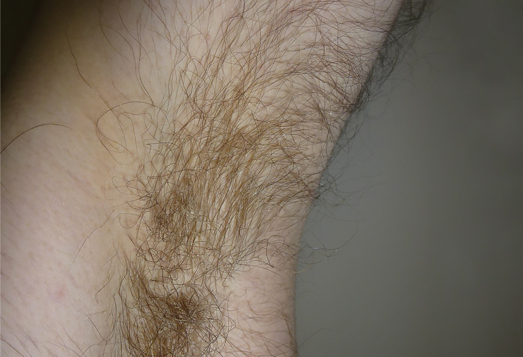
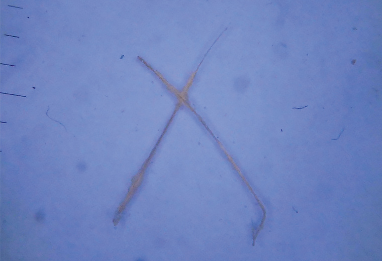
A male aged 15 years presented with a one-month history of malodorous, dirty-looking, yellowish hair over both axillae (Figure 1). The area of affected hair had increased progressively, with associated development of itch and discomfort. Creamy yellow concretions involving several hair shafts were also observed. Polarised contact dermoscopy of pulled hairs showed yellowish and white cotton-like structures surrounding the hair sheath (Figure 2). Affected hairs removed for bacterial and fungal culture and Wood’s lamp study revealed yellowish fluorescence covering the hair shaft. Topical clindamycin 1% twice a day was recommended with good response.

Figure 1. Dyschromia and thickening of the hair of the armpit

Figure 2. Contact dermoscopic image (x10) shows white-yellowish structures forming pods adhered to the hair of the armpit.
Question 1
What is the most likely diagnosis?
Question 2
What differential diagnoses should be considered?
Question 3
What investigations would be most beneficial to confirm the underlying diagnosis?
Question 4
How do you treat this condition?
Answer 1
Trichomycosis axillaris (or trichobacteriosis) is a superficial bacterial infection of the soft keratin of hair located in the armpit, pubis or, less commonly, the scrotum or scalp.1 It is often associated with poor hygiene, obesity and hyperhidrosis.1,2 This disease can stain clothes and may affect social relationships. Trichomycosis axillaris is characterised by white or yellowish nodules on the hair; less frequently, the nodules may be red or black nodules. The nodules are formed by an extremely high number of bacteria.3 The root and the adjacent skin are not usually involved. Although the chemical composition of the cast substance is not yet clear, it is thought to be the metabolic remains of bacteria in the apocrine glands. Corynebacterium spp, Gram‑positive bacterial microorganisms, are the most common ethiopathogical agent involved. Although some cases of Serratia marcescens have been reported, the usual bacteria are C. tenuis, C. propinguum or C. flavescens.2
Trichomycosis axillaris, pitted keratolysis and erythrasma form the corynebacterial infection triad. Pitted keratolysis is not uncommon among athletes and individuals in professions with greater use of occlusive footwear. The lesions tend to be multiple superficial, rounded depressions that measure 0.5–7 mm in diameter and predominantly affect weight-bearing areas of the soles.4 Erythrasma is a superficial infection caused by C. minutissimum and affects the major skin folds and the interdigital regions of the feet. It is characterised by erythematous brown, scaly patches and maceration, and it demonstrates coral-red fluorescence under the Wood's lamp.
Answer 2
The main differential diagnoses include tinea, bromhidrosis, inverse psoriasis, contact dermatitis and antiperspirant residue. Other specific trichological entities that need to be considered include trichorrhexis nodosa and invaginata, pediculosis, monilethrix, pseudomonilethrix and hair casts.
Tinea of the axilla may also cause substantial discomfort but usually presents as an annular lesion with erythematous and elevated margins. Diagnosis of superficial mycoses can be confirmed by a modified Chicago sky blue stain and potassium hydroxide mount.5
Bromhidrosis is characterised by sweat that has an offensive odour. Rather than follicular involvement, excessive secretions from either apocrine or eccrine glands become malodorous on bacterial breakdown.6
Inverse/intertriginous psoriasis occurs in the axillae areas and is often underdiagnosed and undertreated. It is characterised by erythematous and mild scaly plaques involving submammary, axillary and inguinal regions.
Contact dermatitis presents with pruritic papules and vesicles, erythema and crusts. In these cases, it is important to search for the presence of an associated offending allergen. Follicular stem affectation is uncommon with allergic contact dermatitis.
The presence of antiperspirant residue in axillae may simulate clinical presentation of trichomycosis but is cured by swabbing with 70% ethyl or isopropyl alcohol.
Answer 3
The clinical appearance of trichomycosis axillaris is very characteristic. Commercial testing systems (API-Coryne; BioMèrieux, Marcy-l’Étoile, France) allow easy identification of the causative bacteria in 91% of cases.2 These tests are practical, easy to handle, fast (24-hour) and a useful system for the identification of Gram-negative non-fermentative bacilli, as conventional manual methods take longer and require many biochemical, enzymatic and physiological tests that are not commonly available in laboratories.
Culture of affected hair is an alternative test with high specificity. In this case, bacterial culture media are used, as Sabouraud agar growth is usually negative. However, although capable of providing a confirmatory diagnosis, culturing is slow and complex, as Corynebacterium spp grow with difficulty; there is also a high rate of false negatives when using a bacterial culture.
Although dermoscopy is not well described in the literature, the image observed is helpful: a sheath around the hair composed of cottony structures (Figure 2). Hence, from a clinical perspective, dermoscopy could be an important aid in diagnosis. Feather sign and brush sign have been described previously, although concretions with the appearance of a rosary of crystalline stones may be the most typical features.7
Fluorescence is yellowish-green with Wood's lamp but may be confused with fungal infections.
Answer 4
Hair removal, good hygiene and topical 1% clindamycin, 2% erythromycin or 5% benzoyl peroxide are the treatments most often recommended for trichomycosis axillaris. These agents may also treat coexistent erythrasma.8 As trichomycosis axillarisis is at times associated with hyperhidrosis, the use of antiperspirants, iontophoresis, botulinum toxin or thoracic sympathectomy could also be useful. Topical ketoconazole or systemic itraconazole have been found to be effective.
Key points
- Trichomycosis axillaris (or trichobacteriosis) is a relatively common bacterial infection that affects axillary (and sometimes pubic) hair and is caused by bacteria of the genus Corynebacterium.
- Prevalence increases in countries with high humidity and high temperature climates.
- The characteristic clinical presentation and clear response to treatment assists the diagnosis. The presence of a yellowish substance that wraps the hair without invading the cortex on dermoscopy, and positive fluorescence with Wood’s lamp, help to confirm the diagnosis.
- The recommended treatment includes shaving the affected area, good hygiene, topical antibiotics (eg erythromycin or clindamycin) to prevent recurrence, and 5% benzoyl peroxide.