With an increased emphasis on awareness and diagnosis of concussion in both professional and amateur sports, the frequency of reported concussions has significantly increased over the past decade. Australian data suggest that the frequency of reported concussions increased by 60.5% between 2002 and 2011, leading to an annual incidence of >4500 hospitalisations from sporting events.1,2 The costs associated with hospitalisation from these incidents was approximately $18 million, representing a significant burden on the health system.2 The majority of traumatic brain injuries occur in patients aged 15–64 years during participation in sports.1 Although most cases resolve spontaneously without medical intervention over a period of 7–10 days, a subset of patients will report ongoing symptoms.3 Optimal diagnosis and, where indicated, treatment is therefore essential to reduce the impact on either school, work or daily activities and subsequent quality of life.4
Concussion is a diffuse brain injury. As such, signs and symptoms on presentation may be both complex and highly variable. Immediately following impact, the patient may have confusion, dizziness and amnesia. Additional early symptoms include headaches, difficulty concentrating on tasks, sensitivity to light and visual disturbances.5 Despite previous misconceptions, concussion often does not involve the loss of consciousness, which is reported in <10% of cases.6
The widely endorsed Sports Concussion Assessment Tool 5th Edition (SCAT5) and Concussion Recognition Tool (CRT) have shown high sensitivity and specificity in detecting concussion at the sports arena. However, the use of these tests is often restricted to professional or semi-professional sporting events, where trained medical professionals are more readily available.7,8 Accordingly, it has been reported that up to 90% of players may not be aware of a potential concussion diagnosis and go unassessed by health professionals.9 This presents particular difficulties for general practitioners (GPs), who will often see patients at a time after the concussive event. Differentiation of non-specific concussion symptoms from pre-existing conditions is a further confounder.
As more than half of the brain’s pathways are dedicated to vision and eye movement control, a diffuse brain injury such as concussion will often affect the visual system.10 Research has found that visual disorders may occur in up to 69% of patients following mild traumatic brain injuries.11 For children, adolescents and young adults, this may have a detrimental effect on their ability to complete academic activities. In adults, this may lead to difficulties performing crucial daily activities such as driving and coping with work. Early impact or sideline tests include the question, ‘Have you noticed blurred or double vision?’; however, the potential impact on the visual system following concussion may require more in-depth clinical examination and investigation. This literature review aims to inform GPs about the ocular defects associated with concussion and to highlight the GPs’ important role within the concussion paradigm.
Methods
A PubMed search using Medical Subject Headings (MeSH) terms including ‘brain concussion’, ‘concussion’, ‘vision’, ‘eye movement’ or ‘visual dysfunction’ identified 495 papers. After screening for duplicates, this was reduced to 220 papers. These were screened individually; only peer-reviewed, human-based studies published within the past 10 years were considered. Of these papers, only those that tested oculomotor dysfunction following a concussion were included in this analysis (15 papers). This search was conducted on 10 May 2019.
Oculomotor dysfunction
Studies in both adolescent and adult patients have described persistent clinical features affecting the visual system following a concussion (Table 1). These may include diplopia (double vision), decreased visual attention, abnormal pupils and an increase in abnormal eye tracking movements.12 However, the literature in this area is surprisingly limited. The primary research focus has been on the more obvious cognitive and neuropsychological changes immediately following brain trauma. A recent study of military personnel with a past history of mild traumatic brain injury (mTBI) identified a significant relationship between visual complaints and concussion, even five years post-injury.13 This suggests that clinicians must remain aware of possible visual dysfunction years after the event, particularly in more traumatic cases. The incidence of visual symptoms appears to remain relatively consistent across mTBI of different causes and intensities.14
| Table 1. Symptoms and signs affecting the visual system following a concussion |
| Symptoms |
Signs |
- Loss of vision
- Blurred vision
- Double vision
- Reading difficulties
- Photophobia
- Headaches with visual tasks
- Difficulty tracking fast objects
|
- Reduced distance or reading acuity
- Anisocoria
- Impaired vestibulo-ocular reflex
- Slowed or reduced eye movements
- Impaired convergence
- Impaired accommodation
- Decreased visual attention
|
Accommodation
Accommodation refers to a shift in the focus of the eye to maintain a clear image of an object as it varies its distance from the observer, primarily as it approaches. The neural control of accommodation requires afferent input to the visual cortex, and brainstem control of eye movements and the ciliary muscle. The complexity of this pathway makes it potentially susceptible to damage, with resultant dysfunction after a concussion. Within the clinical setting, this may manifest as complaints of blurred vision, asthenopia (tired eyes) and difficulty reading.15
Master et al showed reduced accommodative amplitudes in 51% of young patients with concussion when compared with age-matched normative values.11 Similarly, Capo-Aponte et al prospectively examined 20 military personnel following mTBI and reported reduced amplitudes of accommodation and accommodative facility in 65% and 35% of patients, respectively.12
Accommodative capacity is age dependent. Healthy young or adolescent patients can maintain clear focus on near objects (as close as <10 cm); however, this capacity progressively reduces with age as the crystalline lens gradually becomes more rigid. Therefore, younger patients may be more affected by accommodation dysfunction of neurological origin than older patients. The level of accommodation can also vary with refractive error such as myopia, in which accommodative lag is commonly reported; therefore, a history of wearing glasses or using contact lenses is important adjunctive information.
Accommodation is tested by slowly bringing text towards the subject until the point of blur. This distance is then matched against age-related norms. Specialist equipment (Royal Air Force [RAF] Rule) is designed to measure accommodative amplitudes within ophthalmic practices. However, in the general practice setting, a finding of gross accommodation or a significant reduction through repeated measures – in conjunction with symptoms including difficulty concentrating, headaches following a relatively short period of near work or the appearance of jumbled words when reading – would necessitate referral to a neuro-ophthalmologist for further measurement and possible rehabilitation.
Convergence
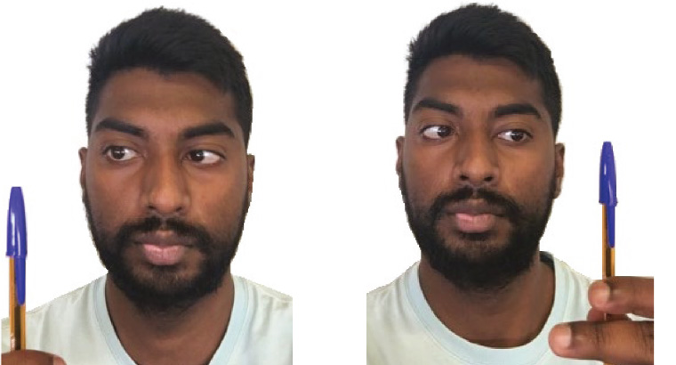
Convergence is the simultaneous movement of both eyes inwards to maintain binocular fixation on a target (Figure 1). Cortical areas including the visual cortex, parietal lobe, frontal eye fields, supra-oculomotor area and cerebellum are involved in the convergence pathway.16 This neurological pathway works closely with accommodation to provide concurrent clear focus on near objects and is highly susceptible to damage by concussion. Convergence abnormalities are reported in 14–55% of athletes,17–19 48–55% of military personnel11,20 and 14–49% of outpatient groups11,21,22 previously diagnosed with concussion. Poor convergence will manifest clinically with double vision following near work, and blurred vision or asthenopia with prolonged near tasks such as reading.
 Figure 1. Eyes before (left) and after (right) converging on a pen. Convergence is tested by measuring the convergence near point.
Up to 40% of patients have ongoing visual symptoms suggestive of convergence dysfunction for at least one month following the concussive episode.23 This suggests high visual morbidity, potentially causing significant disruption to reading and computer work. For athletes, this may affect performance such as their ability to track a ball. For military or security personnel, these symptoms could be particularly dangerous when undertaking rapid-response activities.
Convergence is tested by slowly bringing a pen (or target) closer to the patient’s eyes (Figure 1). The patient is asked to maintain a single image and warned that the target may become blurred as it is brought forward (because of accommodative ability). Clinicians should look for a break in binocular fusion, which would manifest with an obvious divergence of one or both eyes from the target and/or the patient reporting diplopia. It is important to note that approximately 0.2% of the general population will have an abnormal convergence near point, clinically referred to as convergence insufficiency.24 A diagnosis of clinically-reduced convergence may indicate a range of interconnected conditions of the oculomotor system.25 Therefore, referral to an ophthalmologist or orthoptist is recommended.
Saccades
Saccades are rapid eye movements between two or more separate fixation points, at any angular position. Types of saccades are listed in Table 2. The saccadic pathway also involves multiple cortical, cerebellar and brainstem control areas, again making it highly vulnerable to damage following mTBI. Saccadic oculomotor impairments have been linked to both structural and functional abnormalities in the parietal, frontal and temporal areas, and in the caudate.26
|
Table 2. Types of saccades
|
|
Type of saccade
|
Definition
|
|
Visually guided saccade
|
Eyes move toward a visual stimulus
|
|
Anti-saccade
|
Eyes move away from visual stimulus
|
|
Memory-guided saccade
|
Eyes move toward a remembered location
|
|
Predictive saccade
|
Eyes are kept on a target moving in a predictive spatial manner
|
High rates of saccadic dysfunction – which affects up to 30% of patients with a concussion – have been reported in the literature.11,12,20 One study found that patients with a concussion needed to make more saccadic eye movements to complete a number-naming test, when compared with controls who did not have a concussion.27 Patients with an acute concussion have been found to show significant impairment when doing higher cortical saccadic tasks, such as memory-guided saccades and anti-saccades.28 Evidence suggests that individuals with persistent post-concussive symptoms are more likely to have saccadic dysfunction when compared with patients who had a faster recovery.29
Saccadic dysfunction manifests with increased latencies (time between the target presentation and saccade initiation) and dysmetria (lack of coordination of eye movements).28,29 Practically, patients may find it challenging to read text, or complain of difficulty scanning the environment while driving.
Saccadic function (congruency and accuracy) can be readily tested by observing the patient move their eyes on command between two close targets (Figure 2). Poor saccadic accuracy requiring a secondary eye movement adjustment to the target may be seen by the clinician. In the case of significant dysfunction, the patient should be asked to abstain from critical tasks such as driving and operating machinery. Specialist referral for additional neurological deficits may be necessary at this point.
The King-Devick test has been used on the sidelines and during recovery following concussion episodes. Players are asked to read aloud a set of numbers across a page as quickly as possible. This tool primarily measures saccades; however, it also assesses cognition and attention faculties.30 The test can be compared with a baseline finding if available, or to age-appropriate normative values. The King-Devick test has been increasingly used within general practice offices, particularly those with sporting associations, for diagnostic purposes and to track recovery as part of a return-to-play protocol.

Figure 2. For horizontal saccade testing, patients fixate continuously between a target on their right and left. The examiner observes for the complete and simultaneous movement of both eyes to each target. The same can be applied for vertical saccade testing, with a pen held high and low.
Smooth pursuit eye movements
Smooth pursuit eye movements are performed to closely track a moving target, holding the image of the moving target steady on the fovea. As with other types of ocular movement, smooth pursuit requires the integration of multiple cortical areas and is similarly susceptible to concussion.
Few papers discuss concussion-related smooth pursuit dysfunction. However, those that exist report high rates of abnormalities.12,20,31 Within the clinical setting, this would result in increased difficulty following moving objects, such as when catching a ball and tracking nearby cars while driving. Examining smooth pursuits can be achieved by asking the patient to track a pen across the vertical and horizontal planes. Jerky movements or failure to adequately follow the target may indicate problems with smooth pursuit movements. Even with extensive ophthalmological training, assessing subtle defects in smooth pursuit eye movements can be challenging in clinical practice. Specialist referral is suggested if smooth pursuit or tracking dysfunction is suspected.
In the research setting, using video-oculography to track disconjugate eye movements is gaining popularity as a possible marker for concussion.32 Maruta et al examined a population with chronic concussive symptoms and found a significant decline in their predictive visual tracking capabilities.33,34 Using diffusion tensor imaging, researchers attributed these oculomotor impairments to white matter tract damage in the corona radiata, left superior cerebellar peduncle and genu of the corpus collosum. These patients also showed reduced attention and working memory, highlighting the potential need for multidisciplinary referral to both ophthalmologists and neurologists for more extensive examination. A greater understanding of video-oculography metrics and the ability to provide a portable, readily accessible model will help facilitate translation of this technology into the clinic.35
Other conditions
Visual field anomalies have been reported following significant trauma but are rare following sports-related concussive episodes.14,35 Visual field defects related to significant trauma include homonymous hemianopia and quadrantanopia. A confrontation field test can be performed in the clinic if field loss is suspected, with referral for formal visual field testing if there are any concerns.
Cranial nerve palsy may also occur following severe trauma.14 The patient would present with a manifest strabismus, and report diplopia.
Visual field loss and cranial neuropathy are not expected in concussion and should prompt further neurological examination and neuroimaging for a structural pathology.
Oculomotor rehabilitation
Although research is quite limited, there is some evidence supporting vision therapy/exercises as an effective strategy for rehabilitation.36,21 Current protocols in Australian sport recommend rest and a step-wise return-to-play guideline.37 However, recovery will be dependent on many factors including the number and severity of the existing ocular defects. Ciuffreda et al found that treatment in the form of eye tracking, accommodation and convergence exercises may expedite recovery and completely alleviate symptoms in almost 90% of patients with mTBI who have persistent oculomotor defects.36 This can require between 10 and 30 vision therapy sessions over an eight-month period until symptoms resolve.36
Conclusion
Many forms of ocular dysfunction are found in acute and chronic cases of concussion. Being the primary medical contact for many patients with a concussion, either in the immediate or short-term period following the initial trauma, GPs need to be vigilant about the ocular dysfunction, as well as the behavioural, physical and emotional symptoms associated with concussion. Practical tips for assessment of ocular dysfunction following concussion are provided in Box 1. If the GP suspects visual and oculomotor impairment, referral to an ophthalmologist will provide patients with a complete ocular assessment and earlier implementation of treatment and rehabilitation.
| Box 1. Practical tips for concussion assessments |
- Subtle oculomotor dysfunction may be described by the patient as:
- increased difficulty when completing near tasks
- headache and eye strain after shorter duration of visual effort.
- Standard assessment of extra-ocular muscles and pupillary response can be supplemented with tests for:
- convergence
- accommodation
- saccadic eye movement and smooth pursuits.
- Signs and symptoms can persist in some patients for weeks or months after concussion.
- Visual field loss and cranial nerve palsies are rare in concussion and should be investigated further.
|
Key points
- GPs must be alert to persistent oculomotor dysfunction following concussion including impairments to accommodation, convergence, smooth pursuits and saccadic eye movements.
- Developing strong relationships between GPs and ophthalmologists is important to provide the best evidence-based care for patients with concussion-related visual dysfunction.
- Patients with persistent visual disturbances may benefit from oculomotor rehabilitation, such as convergence, accommodation and eye tracking exercises.