Case
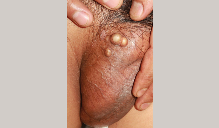
A man aged 45 years with no notable medical history presented with a one-year history of asymptomatic scrotal nodules. He had a history of progressively enlarging scrotal lesions with no preceding local trauma. He had no other lesions on the shaft or glans, no urethral or urinary symptoms and he denied a previous history of unsafe sexual practices. Physical examination revealed three whitish/skin-coloured nodules of 0.5 cm each without local reaction (Figure 1). A hard, solid, homogenous content was noticeable by touch. Laboratory tests revealed normal phosphorus and calcium levels.

Figure 1. Scrotal calcinosis; three indurated whitish/skin-coloured nodules located at right scrotum
Question 1
What is the diagnosis?
Question 2
What are the potential differential diagnoses?
Question 3
What are the treatment options for this condition?
Answer 1
This condition is known as scrotal calcinosis (formerly idiopathic scrotal calcinosis).1 It is a disorder characterised by the progressive appearance of solitary or multiple calcified, painless nodules on the scrotum. The nodules usually appear during early adulthood and tend to increase in size and number with age. They are usually asymptomatic but can be pruritic or result in a heavy sensation or chalky material discharge.2
Scrotal calcinosis was considered idiopathic in the past because no association with other lesions or known systemic pre-conditions was observed. It is not related to a calcium phosphate imbalance or renal insufficiency. Infiltration of foreign body material or previous local trauma as predisposing factors remain controversial.3
The condition has more recently been attributed to dystrophic calcification of epidermoid cysts.4 It occurs as a process of subclinical inflammation, rupture, dystrophic calcification and finally obliteration of the cyst wall.5
Answer 2
Scrotal calcinosis is easily recognised if suspected. The patient presented with multiple epidermal cystic lesions characterised by their hard homogenous content, with a solid appearance. Epidermoid carcinoma of the scrotum is very rare and usually manifests as a single verrucous indurated mass. Testicular cancer is deeper and does not have epidermal involvement.
Scrotal calcinosis can be confused or coexist with typical epidermoid cysts located at the scrotum or nearby areas.6
It lacks the inflammatory appearance of furuncles or abscesses and rarely develops superinfection.
Answer 3
Scrotal calcinosis is a benign condition, and surgical intervention is only recommended in severe cases with disturbance of scrotal appearance or in the context of associated symptoms. Surgical excision is the main procedure. Because of the laxity of scrotal skin, a single-stage excision can be easily performed for small or medium defects.2 A recent novel technique using an erbium:YAG laser has been trialled as a less invasive method. It has good aesthetic and functional results with lower risks of injury and infection.7
Case continued
The patient was reassured about the benign nature of his condition. He had no symptoms or aesthetic concerns and it did not interfere with his sex life. No further intervention was required.
Key points
- Scrotal calcinosis is a benign condition that can be easily recognised if suspected.
- Surgical treatment is only recommended in the context of local symptoms or aesthetic reasons.