Case
A man aged 75 years presented with concerns after finding bright red blood stains in his underwear. This had been occurring intermittently for the past few months, but had intensified over the last week. He reported no clots. He stated he otherwise felt well and specifically reported no pain. He passed motions regularly without difficulty, denied any blood in his stools and stated he had never had haemorrhoids. He had seen no blood in his urine. He denied epistaxis or bleeding from other areas. There was no unexplained bruising. He was on no antiplatelet or anticoagulant therapy.
A rectal exam was performed in order to exclude a possible rectal origin, and the results were unremarkable. Skin changes to his scrotum were noticed during this examination. Closer examination of the scrotum identified a widespread rash consisting of vasculitic papules – both individual and in clusters (Figure 1). These were subsequently considered to be the origin of the bleeding.
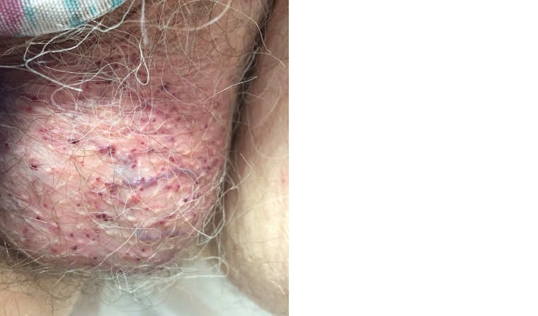
Figure 1. The vasculitic papular rash found on examination of the scrotum
Question 1
What is this condition?
Question 2
What is the typical clinical appearance and distribution of this condition?
Question 3
How prevalent is this condition?
Question 4
What is the pathogenesis of this condition?
Question 5
What are the different variants? How are the different variants distinguished?
Question 6
What are the management options?
Question 7
What differentials could be considered if there was uncertainty about the diagnosis?
Answer 1
The scrotal changes were consistent with angiokeratoma of Fordyce.1
Answer 2
Angiokeratomas are keratotic, vascular skin changes of dark violet to black appearance that arise as individual papules or in clusters.2 They are firm to palpation and generally do not blanch on compression.2
Angiokeratoma of Fordyce classically refers to these changes when found on the scrotum,3 although it can also refer to angiokeratomas on the labia majora, inner thigh and lower abdomen.2,3 They tend to be symptomless and are often only noticed after they bleed from minor trauma, such as intercourse.2,3
Answer 3
This condition is much more common in men and has an increasing prevalence with age, affecting 0.6% of males aged 16 years and rising to 16.7% of men over 70 years of age.4
Answer 4
While the exact pathogenesis of this is unclear, angiokeratomas result from vessel dilation and damaged vascular endothelial cells,2 and there appears to be an association with increased venous pressure.4
Answer 5
There are generally considered to be four other forms of angiokeratoma. These can be subdivided into localised and systemic forms.2,3
In addition to angiokeratoma of Fordyce, the localised forms are:
- angiokeratoma circumscriptum – unilateral changes affecting one of the lower limbs or trunk; found at birth, they can become quite large
- angiokeratoma Mibelli – lesions affecting the dorsal surfaces of the hands and feet; they tend to arise during childhood or adolescence predominantly, but not exclusively, in young girls
- sporadic angiokeratoma – clinically resemble the Mibelli type and are most commonly found on the legs, although they can affect any part of the body; they tend to arise between the second and fourth decades.
The systemic form is angiokeratoma corporis diffusum. This manifests in the context of Fabry disease, a rare, serious, recessive X-linked condition that results in a deficiency of lysosomal enzyme alpha-galactosidase A, which leads to an accumulation of glycolipid in many tissues, including skin, heart and kidneys.5 There is a subsequent broad scope of potential complications, including renal failure, cardiomyopathy, gastrointestinal distress and neuropathy.5 The vascular lesions associated with this condition occur on the trunk and proximal limbs and manifest before puberty.5
Answer 6
As described, angiokeratoma corporis diffusum is a serious genetic condition with the potential for multiple-organ involvement because of the widespread inappropriate glycolipid deposition. As this condition benefits from early intervention, a high suspicion should be maintained when widespread angiokeratomas are found, with early subsequent specialist referral.
Otherwise, angiokeratomas are generally harmless lesions that do not mandate intervention.1,2 However, if there is bleeding or they are of cosmetic concern, options can include cryotherapy, laser therapy, electro cautery or excision.1,2
Answer 7
Some presentations of angiokeratoma of Fordyce may appear similar to a range of other pigmented lesions, including:
- melanocytic nevi
- malignant melanoma
- verruca vulgaris
- condylomata acuminatum
- verrucous hemangiomas
- hereditary hemorrhagic telangiectasia
- lymphangioma circumscriptum
- pigmented basal cell carcinoma
- Spitz nevi
- seborrheic keratosis
- dermatofibromas
- pyogenic granuloma.6
Biopsy and/or referral to a dermatologist should be considered if there is doubt about the diagnosis.
In summary
- Angiokeratoma of Fordyce is considered a benign condition.
- Intervention is only required if there is bleeding or cosmetic concerns.
- Widespread angiokeratomas should raise suspicion for the serious genetic condition Fabry disease, which warrants urgent specialist opinion if suspected.