Scuba diving, during which a diver uses a self-contained underwater breathing apparatus (scuba), is a popular recreational activity resulting in frequent presentations to the general practitioner (GP). Incorrect diagnosis and management of otological diving injuries can lead to significant morbidity including chronic vestibulopathy and hearing loss. As many patients present to the GP with diving-related concerns, it is pertinent that treating clinicians are aware of the common presentations, initial treatment and prevention of such injuries.
Epidemiology
Diving-related otological injuries account for 65–72% of all diving-related presentations to practitioners.1,2 Middle ear barotrauma (MEB) is most common, accounting for nearly 50% of presentations.2 Decompression sickness (DCS) is one of the most severe diving-related complications and has an Australian incidence rate of 10 per 100,000 dives.3 Inner ear DCS (IEDCS) is rare, with an estimated recreational diving incidence rate of 0.01–0.03%.4
Pathophysiology – A lesson in physics
Otological diving injuries occur as a result of the effect of ambient pressure changes on the ear structures. At sea level the atmospheric pressure is 1 atmosphere (atm). For every 10 m a diver descends below this level, the ambient pressure increases by 1 atm. For most purposes, atmospheric gas is made up of 21% oxygen and 79% nitrogen, similar to compressed gases found in a scuba tank.
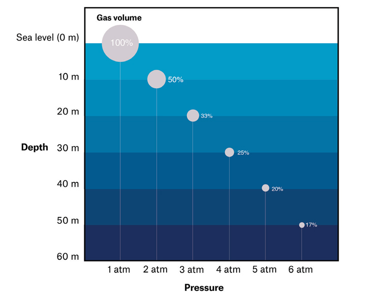
Two important laws of physics govern the physiology associated with pressure changes during scuba diving. Boyle’s Law (P1V1 = P2V2) dictates that the pressure and volume of a gas at a constant temperature are inversely proportional. As the diver descends, ambient pressure increases. The middle ear, being an air-filled space, correspondingly reduces its volume by 50% in the first 10 m of descent (Figure 1). If equalisation does not occur, then a vacuum is created, placing pressure on the tympanic membrane and round and oval windows (the flexible walls of this space). This accounts for direct pressure–related ear injuries during diving.

Figure 1. Boyle’s law dictates that as pressure increases below the water surface, the volume of closed spaces decreases.
As ambient pressure increases, so does the partial pressure of each of the gases within the environment. Henry’s Law dictates that as the partial pressure of a gas increases, a greater amount becomes dissolved in surrounding liquids. During uncontrolled ascent, the reduction in pressure causes release of dissolved gas nitrogen bubbles within tissues or blood that can lead to cell injury and hypoxia, causing the symptoms of DCS.
External ear pathology
Otitis externa
Otitis externa is common among those who spend considerable amounts of time submerged underwater. Prolonged exposure of the ears to water results in a change in canal pH and skin maceration, damaging the primary skin barrier and promoting infection.5 Pseudomonas aeruginosa is the organism most responsible for acute otitis externa following diving.6 The principles of treatment remain the same as for non-divers, with dry ear precautions, aural toilet and topical antimicrobial therapy.
Exostoses
Exostoses are bony outgrowths of the medial ear canal. They commonly occur in divers in response to chronic cold water or wind exposure that triggers refrigeration periosteitis.7 Bony canal stenosis can result in water and wax retention, predisposing to otitis externa. When severe, complete occlusion of the canal results in a conductive hearing loss. Furthermore, unilateral occlusion can lead to an asymmetric caloric stimulus underwater, leading to vertigo.8 Promoting the use of appropriately fitted wetsuit hoods can help to prevent this progression. Referral to an otolaryngologist for consideration of a canalplasty may be necessary in severe or symptomatic cases.
External canal barotrauma
External canal barotrauma can occur as a result of occlusion of the ear canal by exostoses, impacted cerumen and tight wetsuit hoods. An air-filled space between the occlusion and tympanic membrane is created, and negative pressure is generated on descent, leading to swelling and haemorrhagic blistering of the canal wall. Treatment consists of analgesia and topical steroid ear drops.9 External canal barotrauma can be reduced by avoiding tightly fitted hoods and cotton bud use for wax removal. Aural toilet is recommended for persistent wax impaction.
Middle ear pathology
Middle ear barotrauma
MEB occurs when the diver cannot or does not regularly equalise on descent or ascent. This may be due to inexperience or eustachian tube dysfunction (ETD) secondary to rhinosinusitis, allergy or nasopharyngeal obstruction. As external pressures rise on descent, middle ear volumes decrease, creating a vacuum. Serous or haemorrhagic effusion and tympanic membrane rupture can result secondary to the negative pressure.10 Conversely, during ascent, middle ear volume expands as pressure decreases. Normally this is passively released through a functional eustachian tube. However, with ETD, volume expansion can create bulging of the tympanic membrane and perforation.
Symptoms include dullness or pain in the ear and hearing loss. Tympanic membrane perforation during a dive can cause severe vertigo from caloric stimulation of cold water in the middle ear. If nausea and vomiting ensues, this can be fatal because of obligate mouth breathing during scuba diving.11 MEB can be graded using the modified Teed classification,12 which is useful in guiding flying and diving restrictions post-injury (Table 1).
| Table 1. Modified Teed classification12 for middle ear barotrauma* |
| Grading |
Otoscopic findings |
Estimated return to dive |
| Grade 0 |
Normal tympanic membrane |
Seven to 10 days (complete resolution) |
| Grade 1 |
Tympanic membrane erythematous/inflamed |
| Grade 3 |
Gross haemorrhage of the tympanic membrane |
Six weeks (blood reabsorption) |
| Grade 4 |
Extensive free blood in middle ear with bubbles visible behind tympanic membrane (haemotympanum) |
| Grade 5 |
Perforation of the tympanic membrane |
Three months (healed perforation) |
| *Grade 2 has not been included in this modification |
Management involves exclusion of inner ear barotrauma, short-term use of nasal decongestants and/or intranasal steroid sprays and antibiotics for any secondary infection.11 Diving should not recommence until all symptoms have resolved, tympanic membrane perforations have healed and equalisation is possible. In cases of persistent tympanic membrane perforation, surgical repair may be necessary.
Inner ear pathology
Inner ear barotrauma
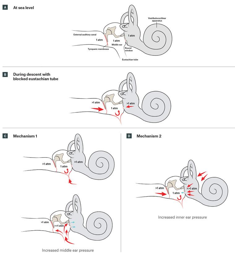
Rupture of the round or oval windows creates a perilymphatic fistula (PLF), which can cause sensorineural hearing loss (SNHL), vertigo and tinnitus. Two mechanisms have been postulated (Figure 2).13,14

Figure 2. Inner ear barotrauma. Click here to enlarge
A. The inner ear at sea level; B. The inner ear during descent with blocked eustachian tube; C. Mechanism 1 suggests a forced Valsalva manoeuvre against a blocked eustachian tube with sudden opening and rush of air into the middle ear. This pressure can cause stapes footplate dislocation and implosion of the oval or round window; D. Mechanism 2 suggests a forced Valsalva manoeuvre against a blocked eustachian tube with increased cerebrospinal fluid pressures transmitted through the cochlear aqueduct and otic fluids leading to round window explosion.
Patients may present with an unsteady gait and ear examination findings similar to MEB, though the ear can also appear completely normal. Tuning fork assessment can be unreliable, thus formal audiometry is required. Fistula testing by applying abrupt tragal pressure to the affected ear or air insufflation by pneumatic otoscopy will elicit nystagmus if positive. However, a negative fistula sign does not exclude PLF.15
Management for PLF is initially conservative with strict bed rest, head elevation, high-dose oral steroids, stool softeners, anti-emetic medications and avoidance of straining.16 Audiometry should be performed daily to monitor for improvement. Progressive SNHL or worsening vestibular function should prompt urgent referral to an ear, nose and throat (ENT) surgeon for surgical repair, which should ideally be performed within 10 days of injury.17 Divers should undergo a fitness to dive assessment by an otolaryngologist or dive medicine physician prior to recommencing diving.8
Inner ear decompression sickness
Two main theories exist for inner ear DCS. The first is formation of inert gas bubbles within the microvasculature or otic fluids of the vestibulocochlear apparatus on ascent. The second is arterial gas emboli shunted from the venous system (ie patent foramen ovale).11,13,18 Patients commonly present with vertigo, nausea and vomiting, although hearing loss and other central nervous system symptoms frequently occur.19 Examination usually reveals a normal external canal and tympanic membrane. Tuning forks may reveal SNHL, and nystagmus is usually present.
It is important for inner ear DCS to be differentiated from inner ear barotrauma as treatment is markedly different (Table 2).20 Treatment of inner ear DCS includes early recompression with hyperbaric oxygen, preferably within five hours of symptom onset. In the general practice setting, it is recommended that administration of 100% oxygen and intravenous saline or oral fluids be provided. Urgent referral to the nearest centre with a hyperbaric chamber is critical to long-term prognosis.4
If there is doubt regarding whether the diagnosis is inner ear barotrauma or DCS, patients should be treated for DCS as it is more severe and can lead to persistent vertigo and ataxia.
| Table 2. Characteristic features differentiating inner ear barotrauma and inner ear decompression sickness |
| Inner ear barotrauma |
Inner ear decompression sickness |
| Conductive or mixed hearing loss |
Sensorineural hearing loss |
| Descent or ascent |
Ascent |
| Cochlear symptoms (ie hearing loss predominates) |
Vestibular symptoms predominant; right sided |
| History of forced or difficult Valsalva manoeuvre |
Not associated with a history of eustachian tube dysfunction |
| Low-risk dive profile |
Dive profile >15 m, technical diving (helium mixtures), multiple dives over a short period |
| Isolated inner ear symptoms |
Other neurological/dermatological manifestations |
Prevention and fitness to dive
Adequate ventilation of the middle ear is essential to the prevention of otological injury. Divers with conditions predisposing them to ETD should avoid diving, and those who are unable to autoinsufflate the ear underwater should immediately abort the dive. Controlling the rate of ascent and taking decompression stops will reduce the risk of DCS. To avoid nitrogen excess, flying is also not recommended within 24 hours following diving.13 The use of oral or topical decongestants is discouraged to treat ETD or sinusitis prior to diving as their effects may wear off while underwater, leaving the diver in danger.13
Fitness to return to diving depends on residual symptoms and ongoing pathology. Persisting neurology, especially affecting the vestibular system, is associated with high risk. An estimated 90% of patients with previous diving-related vestibular dysfunction have ongoing long-term deficits necessitating thorough assessment prior to continuing scuba diving.18 Some authors recommend full vestibulocochlear assessment and exclusion of a right-to-left vascular shunt in those who have previously had inner ear DCS.6,18
Return to diving is possible following otological surgery. Divers should wait three months following tympanoplasty, with diving permissible once the tympanic membrane has healed and function returned.21 Limited evidence exists for return to diving following stapes surgery or canal wall down mastoidectomy and while some authors do advocate this,22,23 in our opinion, the potential lethal risks of experiencing vertigo and vomiting underwater far outweigh any benefits. We recommend that each patient undergoes a full assessment by an otolaryngologist and return to diving is judged on a case-by-case basis and any recommendations are clearly documented.
Conclusion
Patients may present to the GP with a number of injuries related to scuba diving. The ability of the GP to understand the physiology, recognise pathologies and initiate management can greatly influence the patient’s long-term outcomes and recovery. Furthermore, fitness to dive certifications for people with a history of ENT conditions and those who have previously had a dive-related injury are mandatory in Australia.