Case
A Caucasian female, aged 40 years, previously healthy, presented with an eight-week history of a pruritic erythematous skin lesion affecting the inframammary creases. There was no odor or discharge in the area and the patient did not report any systemic symptoms. She had been treated empirically with clotrimazole cream twice a day for the past four weeks, without relief. The patient had a history of overweight and dyslipidaemia and was taking simvastatin. Her family history was significant for type 2 diabetes and psoriasis. Physical examination revealed a well-demarcated, shiny, erythematous plaque with dry scaling, located on the inframammary creases (Figure 1). There were no other physical findings, such as involvement in other skin areas or nail abnormalities.
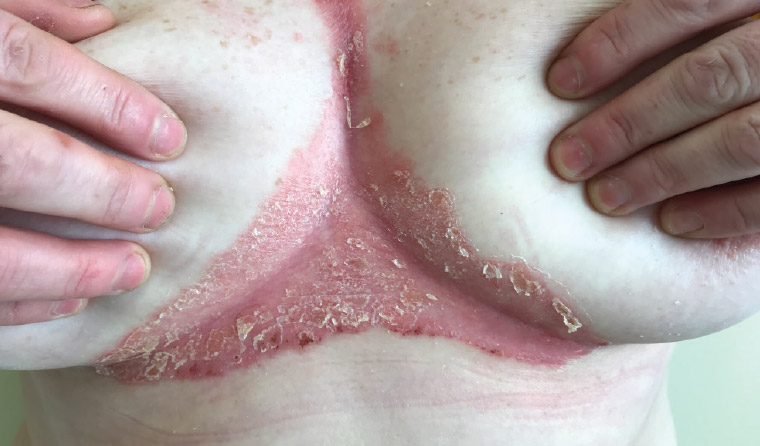
Figure 1. A well-demarcated, shiny, erythematous plaque with dry scaling, located on the inframammary creases
Question 1
What is the most likely diagnosis?
Question 2
What differential diagnosis would you consider?
Question 3
What is the pathogenesis of this phenomenon?
Question 4
How would you treat this patient?
Answer 1
Inverse psoriasis, also called flexural psoriasis, can occur at the point of contact of any two skin folds (intertriginous areas): the inguinal fold, axilla, and external genitalia are the most common locations.1 It is characterised by well-defined, shiny, erythematous plaques, and it usually lacks scale because of the presence of moisture.2 This diagnosis should be considered in all cases of chronic inflammatory dermatitis that affects flexural areas and is unresponsive to antifungal treatment, especially if there is a family history of psoriasis.
Answer 2
Candidal intertrigo is an intertriginous fungal skin infection caused primarily by Candida albicans.3 Lesions usually appear as red plaques with satellite papules and pustules, often associated with a collarette of scale and a foul-smelling odor; pruritus is common, and pain may occur if fissuring or erosion is present.3
Seborrheic dermatitis affects regions rich in sebaceous glands (face, upper chest, axillae and inguinal folds), and it has a multifactorial aetiology that includes individual susceptibility, sebaceous secretions and skin surface fungal colonisation.4 Typically, lesions are distributed symmetrically and present well-delimited erythematous plaques with greasy-looking and yellowish scales of varying extents, causing inflammation, pruritus, and occasionally marked erythema.4
Irritant contact dermatitis is an acute or chronic inflammation of the skin caused by contact with chemical or physical agents.5 Underwire bras can be particular culprits, as the metal wires may cause mechanical trauma and also be a potential source of nickel exposure. In general, during the acute phase, the clinical findings include erythema, scaling, vesiculation and bullae, confined to the area of contact with the irritant; however, diffuse lichenification and fissuring can also occur with chronic disease, causing pain and pruritus.5
Erythrasma is an intertriginous bacterial skin infection caused by Corynebacterium minutissimum.6 Lesions are asymptomatic or minimally pruritic and may manifest as well-defined erythematous to brown patches or as thin plaques, sometimes slightly scaly and wrinkled.6
The possible causes of pruritic erythematous plaques are summarised in Table 1.
| Table 1. Possible causes of pruritic erythematous plaques |
| Condition |
Characteristics of the lesion |
| Candidal intertrigo |
Uniformly erythematous plaques with collarette scaling and satellite papules and pustules; pruritus is common, and a foul‑smelling discharge may be present |
| Erythrasma |
Well-defined erythematous to brown macules or thin plaques, slightly scaly and wrinkled; asymptomatic or minimally pruritic |
| Inverse psoriasis |
Well-demarcated shiny erythematous plaques with minimal to no scale; pruritus is common, and nail abnormalities or a family history of psoriasis can assist diagnosis |
| Irritant contact dermatitis |
Erythematous patches with scaling, vesiculation and bullae in the pattern of the irritant exposure; diffuse lichenification and fissuring can also occur, causing pain and pruritus |
| Seborrheic dermatitis |
Well-delimited erythematous plaques with greasy-looking and yellowish scales of varying extents, causing pruritus and occasionally marked erythema; typically on seborrheic areas |
Answer 3
Inverse psoriasis is considered a specific subtype of psoriasis vulgaris. To date no separate pathogenic mechanism has been found: attempts to prove a correlation with skin microbiota or specific human leukocyte antigen (HLA) subtype have not been sustained.7 Psoriasis is a chronic autoimmune disease with multiple leukocytes and cytokines interacting to produce a sustained immune cell–mediated inflammatory response within the skin itself.1
Answer 4
Low-potency topical steroids are often used as the first-line treatment for the short term, but with a shift to topical vitamin D3 analogues or topical calcineurin inhibitors when the treatment needs to be extended for more than four weeks.8 Side effects of topical steroids tend to occur more frequently with topical steroids of higher potency, in prolonged treatment and on particular areas of the body, such as intertriginous areas, due to thinner skin and increased occlusion.9 Topical calcineurin inhibitors, with modest efficacy in chronic plaque psoriasis, play a bigger role in the treatment of inverse psoriasis.8,10
Key points
- Inverse psoriasis should be considered in all cases of chronic inflammatory dermatitis that affect flexural areas and are unresponsive to antifungal treatment, especially if there is a family background of psoriasis.
- Low-potency topical steroids are often used as the first-line treatment for the short term, but topical calcineurin inhibitors play a bigger role in the treatment of inverse psoriasis.