Case
A woman aged 42 years presented to the hospital with a one-week history of a wrist lesion. It started with a yellow crusted excoriation on her skin after scratching an insect bite. She was treated by her primary care physician, who prescribed mupirocin ointment. After approximately 48 hours, she developed an itchy, tender rash. The patient was understandably concerned and had diligently taken progress photographs (Figure 1). The rash consisted of a well-demarcated, square-shaped geometric erythematous plaque with vesicles, scale and crusting.
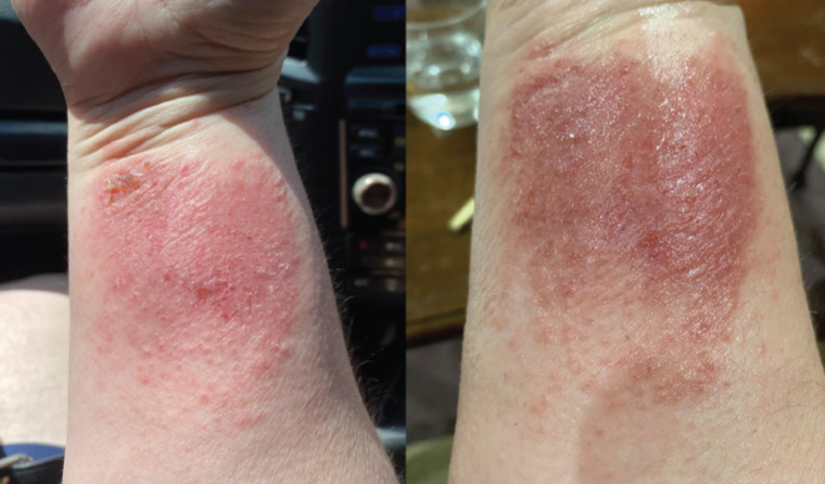
Figure 1. Photographs taken by the patient demonstrate an erythematous plaque on the wrist.
Question 1
What are the differential diagnoses?
Question 2
What further history is important in this case?
Question 3
What initial investigations could assist with making a diagnosis?
Answer 1
Differentials for an erythematous eruption with scale are broad and include several conditions (Table 1). Given the morphology of this lesion and temporal relationship with the application of a new topical therapy, important differentials include irritant and allergic contact dermatitis and infective dermatitis.1
| Table 1. Differentials of an erythematous scaly rash |
| Infective |
Inflammatory |
Malignant |
- Dermatophyte infection (tinea corpis)
- Scabies
|
- Contact dermatitis (irritant or allergic)
- Psoriasis
- Atopic dermatitis
|
|
Answer 2
When suspecting contact dermatitis, it is important to take a careful history that assesses for potential precipitants, including:
- history of presenting illness – compounds/dressing applied to skin, time course of the rash, compromise in skin barrier
- medical history – atopic dermatitis, asthma, allergic rhinitis
- medications – regular medications, allergies to medications and other compounds (eg nickel allergy)
- social history – occupational history with an emphasis on exposure to chemicals and frequent handwashing; pets, hobbies, plants, improvement when not at work
- family history – history of atopy or contact dermatitis.
Answer 3
In this case, a thorough history and examination are sufficient for diagnosis. Laboratory investigations will help to confirm the diagnosis and exclude superimposed conditions. These include:
- skin scrapings for potassium hydroxide fungal microscopy – can exclude zoophilic or geophilic tinea
- bacterial skin swabs for possible superimposed infection
- skin biopsy – could provide support for contact dermatitis and may be used in difficult cases where there is resistance to treatment or no obvious trigger.
Case continued
On further history, the patient reported using mupirocin ointment on the wound for seven days and applying a sanitary pad to absorb the wound exudate. The plaque aligned with the area of the ointment and dressing. She had no history of atopy and no other exposure to new chemicals/materials. The wound was swabbed and had no significant bacterial growth. The patient had no vulval irritation or issues and had been using tampons during menstruation throughout her life without any reaction.
Question 4
What is the difference between irritant and allergic contact dermatitis?
Question 5
What is likely causing her dermatitis, and what would be an appropriate initial management plan?
Question 6
What investigations could provide a definitive diagnosis in this case, and when are they indicated?
Answer 4
Irritant contact dermatitis is caused by direct damage of the skin barrier due to toxic effects of specific substances; the severity of reaction depends on many factors including the type of irritant, duration of exposure, concentration of the irritant and presence of occlusion. Allergic contact dermatitis is caused by a T-cell mediated hypersensitivity reaction to a chemical previously encountered (type IV).2 It can be difficult to distinguish between the two clinically,3 given that both disease processes may be present in certain situations; for example, skin barrier compromise in irritant contact dermatitis can precipitate allergic contact dermatitis to another compound. Overall, irritant dermatitis is much more common when compared with allergic contact dermatitis, and the strict demarcation in this case is a clue that favours this diagnosis.
Answer 5
In this case, either the sanitary pad or the mupirocin are potential culprits. Cases of allergic contact dermatitis have been described to colophony (a common substance used in adhesives and toiletries) in pads4 and to mupirocin.5
Most cases of contact dermatitis can be resolved with cessation of the suspected offending agent. Topical moderate- and high-potency corticosteroids can be used on persistent lesions on the body. An example is methylprednisolone (0.1%) applied once or twice daily for 2–4 weeks.6 Fingertip units are valuable in explaining how much steroid is required: the length of applied product across the fingertip is enough to treat a handprint surface area on the body. Efficacy of topical treatment can be improved by applying steroids under occlusion – the patient can be instructed to apply saline compresses to the affected area after steroid application.
Systemic corticosteroids (up to 25 mg daily, then weaned) can be considered in severe cases where the reaction covers an extensive area of skin (>20%) or involves difficult-to-treat areas such as the hands, face, feet or genitalia. Systemic corticosteroids would not be indicated in this situation given the location and size of the lesion.
Answer 6
Repeat open application test
Repeat application of mupirocin onto a small area would indicate mupirocin sensitisation but would not identify the specific ingredient causing this reaction.
Patch testing
Patch testing is indicated where there is no clear trigger. It will help determine the specific allergen causing the dermatitis to facilitate avoidance for long-term remission. This requires referral to a dermatologist with experience in patch testing. An adhesive containing small areas of labelled allergens is placed onto the back and occluded for 48 hours, with interpretation on day three or four and on day seven. Common allergens include nickel, cosmetics, fragrances and preservatives. Systemic corticosteroids should be weaned prior to investigation, but topical cortisone will not alter the result unless applied on the patch testing site.
There are limitations to patch testing; allergic contact dermatitis may be identified, but patch testing will not provide an adequate answer for irritant contact dermatitis.7 Moreover, positive results may not be relevant – the patient may be positive to substances they are not being exposed to; therefore, specialist interpretation is recommended.
Case continued
The hospital the patient attended did not offer a patch testing service, and the patient was given the options of referral to a public clinic, seeing a dermatologist privately or trialling avoidance of the suspected products. She opted for trial avoidance and treatment with topical steroids; her forearm improved rapidly (Figure 2), and she had no further issues.
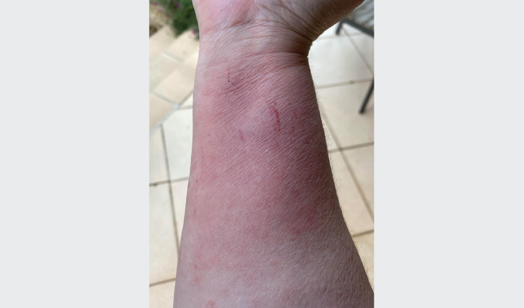
Figure 2. Improvement in the erythematous plaque after avoidance of offending agent and topical treatment
Conclusion
Contact dermatitis is a common dermatological issue in general practice. It may have multiple clinical presentations; when it presents with a well-demarcated erythematous scaly eruption and history of exposure to an offending agent, a clinical diagnosis can be made on the classical contact pattern. Initial management should involve avoidance of the suspected cause and appropriate corticosteroid therapy according to severity.
Key points
- In contact dermatitis, identifying and eliminating the offending agent and applying topical corticosteroids are first-line management options.
- Where multiple potential triggers are identified for contact dermatitis, patch testing with a dermatologist is indicated.
- General skincare measures are valuable adjuncts to treatment and include avoidance of irritants, soap-free washes and the use of emollients for dry skin.