Case
An infant male, aged six months, presented with changes to a lesion on the scalp that had been present since birth. The solitary, yellow–orange, ovoid plaque measured 1 cm × 3 cm and was slightly papular. It had become thicker and more verrucous. The lesion was hairless (Figure 1). There were no similar lesions elsewhere, and the infant was otherwise well.
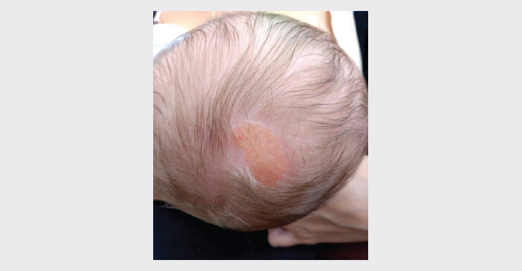
Figure 1. Macroscopic image of scalp lesion
Question 1
What is the most likely diagnosis?
Question 2
How does the diagnosed condition typically present?
Question 3
What differential diagnoses need to be considered?
Question 4
What diagnostic testing is required?
Question 5
What other associated syndromes and complications should be considered?
Question 6
What management is required for this condition?
Answer 1
The lesion is consistent with a sebaceous naevus (also known as a naevus sebaceous of Jadassohn). Considered a type of birthmark, it usually arises on the scalp, but can also appear on the face, neck and forehead. It is a benign hamartoma of the skin. Components of the epidermis become overgrown, including sebaceous glands, hair follicles, apocrine glands and connective tissue, which gives this lesion its classification as a hair follicle tumour.1
Answer 2
A sebaceous naevus will often present in an infant or young child as a solitary, waxy, smooth, and yellow–orange or tan hairless plaque. It is mostly unchanged in infancy due to the quiescence of sebaceous glands. It is usually oval or linear in shape. These lesions can become more pronounced and thicker during adolescence under hormonal influences, appearing bumpy, verrucous or scaly.1 The lesions most commonly occur on the scalp in association with a well-defined, localised area of alopecia.2
Sebaceous naevi occur in approximately 0.3% of newborns,3 and are thought to be caused by postzygotic – that is, not inherited – mosaic mutations in the HRAS or KRAS genes.4
Answer 3
Differential diagnoses of a sebaceous naevus include aplasia cutis congenita, juvenile xanthogranuloma, epidermal naevus, solitary mastocytoma and naevus psiloliparus. The features that distinguish these from a sebaceous naevus are outlined in Table 1.
| Table 1. Features of the differential diagnoses of sebaceous naevus |
| Condition |
Pathophysiology |
Clinical features |
| Aplasia cutis congenita |
A focal absence of skin |
Sharply demarcated, ulcerated plaque covered by a tense epidermal membrane. Re-epithelialisation can result in a scar |
| Juvenile xanthogranuloma |
Rare, benign, proliferative histiocytic disorder of dermal dendrocytes |
Reddish plaques or nodules on the head and neck and upper trunk |
| Epidermal naevus |
Benign hamartomas of the skin |
Present at birth with linear plaques of skin-coloured or brown, verrucous papules that follow the lines of Blaschko, usually on the trunk and limbs |
| Solitary mastocytoma |
Clustering of mast cells |
Red or yellowish plaque or nodule in infancy or early childhood. Lesions can blister or flush if rubbed |
| Naevus psiloliparus |
Mesodermal naevus occurring on the scalp, characterised by paucity of hair and presence of excessive fatty tissue |
Round or oval alopecic patch of the scalp |
Answer 4
The diagnosis of this lesion is usually clinical, its history and appearance often leading to a diagnosis in childhood or adolescence. A diagnostic biopsy will demonstrate the presence of immature hair follicles and sebaceous glands.2 After puberty, the epidermis demonstrates prominent papillomatous hyperplasia with abundant sebaceous glands.
Answer 5
No further evaluation is needed for children presenting with a small, solitary sebaceous naevus. If the lesion is extensive or in the midline, naevus sebaceous syndrome should be considered, and a thorough neurological and ophthalmological examination should be performed and an appropriate specialist referral made. Further skeletal imaging and neuroimaging might also be required. Features of sebaceous naevus syndrome include developmental delay, seizures, hemiparesis or cranial nerve palsies and development of tumours of the eye.5
Although sebaceous naevi tend to remain unchanged throughout life, a variety of benign and malignant epithelial neoplasms might develop within the lesion. Benign tumours include viral warts, trichoblastoma, syringocystadenoma papilliferum, tricholemmoma and sebaceoma.6 Malignant neoplasms include basal cell carcinoma, apocrine carcinoma, trichilemmal carcinoma, sebaceous carcinoma and squamous cell carcinoma.7 Rapid, circumscribed enlargement or ulceration could signal malignant transformation. The incidence and lifetime risk of malignancy remain unclear. Benign and malignant tumours (mainly basal cell carcinomas) are believed to occur in 16% and 8% of cases, respectively.8
Answer 6
Monitoring of a sebaceous naevus would be appropriate initially. If any changes are noted, such as the development of a lump or ulceration within the sebaceous naevus, then a biopsy could be performed. Definitive treatment is a full-thickness excision; however, the necessity and timing of excision to prevent possible future malignancy is unclear.9 Most malignant lesions develop in adulthood, and the frequency of monitoring depends on any changes observed. The decision to excise the lesion should be case based, with consideration of age and the patient’s ability to give consent, extension and location of the lesion, and concerns about the cosmetic appearance and/or risk of malignancy.
Key points
- A sebaceous naevus will often present as an isolated, solitary plaque on the scalp of an infant or child.
- While no acute management is required, monitoring of any changes, or from early adulthood, is advised, given the potential for malignant transformation within these lesions. The frequency would depend on the nature of the changes (if any) observed.
- The optimal timing of definitive surgery will depend on age, lesion features and patient preferences.