Case
A boy aged four months presented with an erythematous and desquamative eruption over intertriginous and flexural sites that had developed one month prior. Skin swabs taken by his general practitioner (GP) were negative for fungal infection. He had been treated with multiple courses of topical corticosteroids with no effect. A detailed history revealed only one relevant exposure: benzalkonium chloride (BAC) in a laundry rinse the family had been using in the general wash for the past six weeks.
The patient had no significant medical history and did not take any regular medications. He had no known medication allergies, and his immunisation status was up to date.
Physical examination revealed well-demarcated annular erythematous plaques associated with desquamation symmetrically distributed over the neck, chest, axillae and antecubital fossae (Figures 1 and 2).
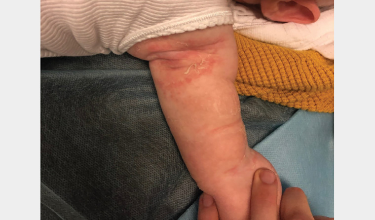
Figure 1. Annular erythematous plaques associated with desquamation over the left axilla and antecubital fossa

Figure 2. Annular erythematous plaques associated with desquamation over the neck and chest
Question 1
What is the most likely diagnosis?
Question 2
What differential diagnoses should be considered?
Answer 1
The most likely diagnosis is granular parakeratosis secondary to BAC exposure. The patient was exposed via his clothing following the addition of laundry rinse (7% BAC) to the household washing machine as a post-wash rinse cycle. Granular parakeratosis is a rare, benign and poorly recognised cutaneous disorder with very distinct clinical features. It classically presents over intertriginous sites as an annular eruption consisting of erythematous to brown papules or plaques associated with marked scaling.1 Lesions can be asymptomatic, pruritic or painful. The incidence of granular parakeratosis is estimated to be 0.005%.2 Although the adult form commonly affects women older than 40 years, granular parakeratosis has been reported in both sexes and patients of all ages.1 Granular parakeratosis is anticipated to be particularly relevant in the context of the COVID-19 pandemic, where the use of antiseptics and disinfectants has undeniably increased. As such, it is expected that cases of granular parakeratosis secondary to BAC exposure will continue to rise.
Answer 2
The list of differential diagnoses for granular parakeratosis is extensive and includes conditions that typically affect intertriginous sites or present with similar clinical findings (Box 1). A broad differential diagnosis should be considered, particularly in the case of a baby aged four months. Relevant differential diagnoses in an infant include irritant or allergic contact dermatitis, dermatophytosis, erythrasma (superficial bacterial infection), flexural psoriasis, Hailey–Hailey disease, streptococcal intertrigo, necrolytic migratory erythema (secondary to glucagonoma) and acrodermatitis enteropathica (secondary to zinc deficiency). It is advisable to take a swab and skin scraping from the area for microscopy, culture and sensitivity testing to rule out an infectious aetiology.
| Box 1. Differential diagnosis of granular parakeratosis1,2 |
- Acanthosis nigricans
- Bowen’s disease
- Confluent and reticulate papillomatosis
- Contact dermatitis – irritant or allergic
- Darier’s disease
- Dermatophytosis
- Dowling–Degos disease
- Epidermal naevus
- Erythrasma
- Hailey–Hailey disease
- Inverse lichen planus
- Post-inflammatory hyperpigmentation
- Seborrhoeic keratosis
- Flexural psoriasis
- Verrucae
- Pemphigus vegetans
|
As a result of the variety and nonspecific nature of clinical presentations that can be seen, diagnosis often relies on the pathognomonic histopathologic findings of granular parakeratosis. The epidermis is acanthotic or psoriasiform with hyperkeratosis, compact parakeratosis and retention of keratohyalin granules in the stratum corneum.1,2
Case continued
The patient was diagnosed with granular parakeratosis secondary to BAC exposure. In this case, the diagnosis was clear based on clinical findings alone, and a biopsy was not required.
Question 3
What is BAC?
Question 4
What is the cause of granular parakeratosis?
Question 5
What is the treatment for granular parakeratosis secondary to BAC exposure?
Answer 3
BAC is a quaternary ammonium compound that acts as a surfactant, causing disruption of cellular lipid bilayers.3,4 It is active against a wide range of bacteria, yeasts and fungi. Although well recognised as a skin irritant, BAC is commonly used as an antiseptic and preservative in skin disinfectants, household and industrial cleaning products, personal care products such as eye drops, and laundry rinse aids. It has been associated with the development of granular parakeratosis in the paediatric population.5
Answer 4
The aetiology of granular parakeratosis is unclear. In granular parakeratosis, there is an alteration of keratinocyte maturation from the stratum granulosum to the stratum corneum, resulting in retention hyperkeratosis.1 This is thought to be caused by a defect in the processing of profilaggrin to filaggrin, leading to retention of keratohyalin granules, elevation of stratum corneum pH and ultimately increased adhesion and defective barrier function.1,5 This process is likely exacerbated by mechanical and chemical irritation, along with occlusive environments.6 It is now speculated that granular parakeratosis may be more of a reactive pattern in response to various stimuli than a distinct clinical disease.1 In recent years, there has been a greater recognition of the association between granular parakeratosis and chemical irritants found in antiseptic products, especially laundry rinse aids and detergents containing BAC.5,6
Answer 5
Treatments including topical and systemic corticosteroids, retinoids, vitamin D analogues, keratolytics and antifungal agents all have variable efficacy.6 Oral antibiotics such as amoxicillin–clavulanic acid may have some benefit.7 General skin cares, including emollients, are recommended. However, these treatments are of limited value if contact with the causative agent is ongoing. Therefore, withdrawal of BAC-containing products is the mainstay of treatment.6 All clothing and linen must be washed in hot water; it can take multiple hot wash cycles to eliminate the irritant. Complete cessation of exposure to BAC usually results in resolution of the rash over 3–4 weeks.5 This condition will often recur with re-exposure to clothes that are still contaminated with BAC. For this reason, many patients experience a course of disease characterised by exacerbations and remissions before complete resolution is seen.
Case continued
The boy’s parents were advised to cease use of the laundry rinse immediately. To prevent recurrence, they were instructed to wash any clothing or linen items that had contact with BAC for at least four cycles before reuse. In the interim, the use of new clothing and linen was recommended if the skin continued to flare. Basic skin cares were advised, such as use of a soap-free wash and regular emollients. After two weeks, complete clearance of the lesions was observed.
Key points
- Granular parakeratosis is poorly recognised and often overlooked as a differential diagnosis of erythematous flexural dermatoses; there exists a limited clinical awareness of its possible precipitants.
- It is important that GPs maintain a high index of suspicion for granular parakeratosis when encountering patients presenting with flexural dermatoses; a history of recent exposure to BAC should specifically be sought.
- Increased awareness of granular parakeratosis could allow for a timelier diagnosis and prevent unnecessary medical visits, investigations and treatments.
- The mainstay of treatment for granular parakeratosis includes identification and removal of products containing BAC in combination with general skin cares.