Case
A man aged 71 years was referred by his general practitioner to a dermatologist for opinion and management of asymptomatic hyperpigmented lesions on his lower limbs. The patient requested referral in order to obtain a specific dermatological diagnosis and to improve the cosmetic appearance of his legs. He noticed the first lesion 10 years prior and had since developed 30–40 similar-appearing lesions, all below the knees. His medical history was remarkable for a 30-year history of poorly controlled non-insulin-dependent (type 2) diabetes, complicated by coronary artery disease, diabetic retinopathy and nephropathy. On examination, the patient had multiple well-demarcated, indented, light brown patches of varying sizes on both lower limbs, with predominance on the anterior aspects (Figures 1,2). There was no involvement above the knees or elsewhere on the body. Otherwise, the skin on his legs showed mild haemosiderin deposition, but there were no varicosities. He did not have evidence of toenail onychomycosis.
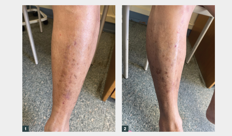
Figure 1. Patient’s left leg.
Figure 2. Patient’s right leg.
Question 1
What is the most likely diagnosis?
Question 2
What are the typical clinical features of the most likely diagnosis?
Question 3
What differential diagnoses should be considered?
Question 4
What diagnostic testing, if any, is indicated?
Question 5
What are the treatment options for diabetic dermopathy?
Answer 1
The most likely diagnosis is diabetic dermopathy.
Answer 2
Diabetic dermopathy presents initially as asymptomatic dull red to pink papules or plaques, predominantly on the pretibial skin, that evolve over the course of several weeks into well-circumscribed, atrophic, brown macules and patches, often with fine scale.1 The lesions may resolve spontaneously over months to years or, more commonly, persist indefinitely. Individuals with diabetic dermopathy typically have long-standing diabetes with microvascular complications. Diabetic dermopathy may affect up to 40% of individuals with diabetes.2
Answer 3
Postinflammatory hyperpigmentation
Postinflammatory hyperpigmentation typically manifests as brown macules
and/or patches that occur as a consequence of an inflammatory dermatosis occurring at the same location/s. Inflammatory dermatoses that often affect the lower limbs and may mimic diabetic dermopathy include psoriasis, lichen planus and atopic dermatitis. A general skin examination may reveal coexisting active lesions typical of the underlying dermatosis.3
Stasis dermatitis
Stasis dermatitis is characterised by scaly, erythematous to hyperpigmented patches occurring on the lower limbs, usually in association with venous disease. It may be differentiated from diabetic dermopathy by the presence of more prominent scale and associated pruritus, as well as the presence of other features of chronic venous insufficiency such as varicosities.4
Pigmented purpuric dermatoses/capillaritis
Pigmented purpuric dermatoses are characterised by petechiae/purpura and haemosiderin deposition, resulting in orange to brown (‘cayenne pepper’) patches. The most common sites of involvement are the lower limbs, although the lesions also may develop at other anatomical sites. The lesions may be asymptomatic or pruritic.
Lichen planus pigmentosus
This rare variant of lichen planus, which is most commonly seen in skin of colour, presents as asymptomatic or mildly pruritic oval or irregularly shaped brown to grey–brown macules and patches.5 It tends to affect sun-exposed sites, but can also involve the lower limbs.6
Answer 4
The diagnosis of diabetic dermopathy is clinical. A skin biopsy is not routinely done but, if performed, shows non-specific findings, such as spongiosis and dermal oedema, red blood cell extravasation and a mild perivascular lymphohistiocytic infiltrate.7 Older lesions show epidermal atrophy and scattered haemosiderin. If a diagnosis of diabetic dermopathy is made, it is prudent to investigate for the microvascular and macrovascular complications of diabetes.
Answer 5
Because diabetic dermopathy is typically asymptomatic and self-resolving, no specific treatment is needed in most cases. Camouflage should be considered for cosmetic purposes8 if desired by the patient. General management should include optimisation of glycaemic control and prevention of the micro- and macrovascular complications of diabetes.9
Key points
- Diabetic dermopathy is a benign, asymptomatic and likely underdiagnosed skin condition seen in up to 40% of individuals with diabetes.
- Diabetic dermopathy is most commonly seen in those with long-standing and/or poorly controlled diabetes with microvascular complications.
- The diagnosis of diabetes is usually known prior to the development of diabetic dermopathy; however, its development may signify the need for tighter glycaemic control and surveillance for other complications of diabetes.