Shoulder pain is a common presenting complaint, representing 1.3% of all general practice presentations, making it the third most common musculoskeletal presentation after spine and knee pathology.1 Patients with chronic (greater than six months’ duration) shoulder pain often experience significant functional disability and reduced psychosocial wellbeing.2
Aim
This article reviews the management of patients presenting with chronic shoulder pain. Key features on history and examination are summarised, and treatment options are discussed.
History and examination
The differential diagnosis for shoulder pain is broad, including both intrinsic, extrinsic and mixed sources of symptoms (Table 1). Rare but serious ‘red flag’ causes of shoulder pain should be considered with a high index of suspicion for patients with a history of cancer or cardiovascular disease, or with risk factors for occult fracture, avascular necrosis or infection (Table 2).
| Table 1. Common causes of chronic shoulder pain and key clinical signs and symptoms3,4 |
| Common pathology |
Signs and symptoms |
| Spinal pain (somatic referred pain or radiculopathy) |
- Dorsal scapular pain
- Pain distal to the elbow
- Reproduction of symptoms with neck movements
|
Instability
- Post-traumatic
- Atraumatic (hereditary multidirectional or overuse acquired)
|
- Younger patient
- Past history of dislocation
- Apprehension of shoulder dislocation or subluxation
- Generalised hypermobility and/or repetitive overuse
|
| Long head bicep and labral pathology |
- Younger to middle-aged patient
- Painful click or mechanical catch
- Pain with O’Brien’s test Position 1 (Figure 1E)
- Pain with Speed’s test (Figure 1F)
- Pain with Yergason’s test
|
| Subacromial pain and minor rotator cuff disease (calcific tendonitis, tendinopathy, partial tear, small to medium full-thickness tear) |
- Middle-aged to older patient
- Painful arc during abduction
- Pain with Hawkins–Kennedy test (Figure 1A)
- Pain with Jobe’s (empty can) test (Figure 1B)
|
| Rotator cuff arthropathy and major rotator disease (large to massive tears) |
- Older patient
- Active versus passive range of motion discrepancy
- Weakness with Jobe’s or drop arm test
- Weakness with external rotation resistance test (Figure 1C)
- Weakness with internal rotation belly press test (Figure 1D)
|
| ACJ arthropathy |
- Patient of any age
- Localised ACJ tenderness
- Pain with O’Brien’s test Positions 1 and 2 (Figure 1E)
|
| Frozen shoulder contracture syndrome |
- Middle-aged patient
- Passive range of motion reduced
|
| Glenohumeral osteoarthritis |
- Middle-aged to older patient
- Passive range of motion reduced
|
| ACJ, acromioclavicular joint. |
| Table 2. Red flag causes of shoulder pain3,4 |
Orthopaedic
- Unreduced dislocation (after past trauma, seizure, electrocution)
- Occult fracture (osteoporosis)
- Avascular necrosis of humeral head (increased risk with steroids, alcohol abuse, previous fracture, sickle cell anaemia)
|
Infection
- Septic arthritis
- Osteomyelitis
|
Inflammatory disease
- Rheumatoid arthritis
- Polymyalgia rheumatica
- Spondyloarthropathy
|
| Malignancy
|
Referred pain from extrinsic location
- Cardiac (acute myocardial infarction, angina)
- Upper gastrointestinal (cholecystitis, pancreatitis, gastro-oesophageal reflux disease)
- Cervical radiculopathy or myelopathy
- Thoracic outlet syndrome (neural, venous, arterial or combined)
|
Demographic factors, past injury and medical history can provide clues towards diagnoses. Younger adults (aged <40 years) should be examined for signs and symptoms of instability due to its much higher prevalence in this age group.5–7 Conversely, the incidence of rotator cuff disease, osteoarthritis and cuff arthropathy is low before early middle age (35–40 years) and steadily increases with age thereafter.8,9 Frozen shoulder contracture syndrome and calcific tendinopathy peak in incidence during middle age (40–70 years) and are over-represented in patients with type 2 diabetes, thyroid dysfunction or dyslipidaemia.10
Eliciting pain site and character is useful. Pain described as originating from the root of the neck with radiation between the shoulder blades or posterior shoulder is more likely cervical in origin. Polymyalgia rheumatica should be considered in bilateral shoulder pain, especially in older adults. Shoulder pathology rarely spreads below the elbow and neurological symptoms do not originate from shoulder conditions.
The minimum standard examination should include neurovascular screening, palpation of soft tissues and bony prominences (not forgetting the sternoclavicular joint and axilla), followed by active and passive movements of the neck and shoulder. The examination can be augmented by numerous special tests (Figure 1). No single test has adequate reliability and accuracy to form a diagnosis in isolation, and therefore tests should be performed in clusters and considered alongside a patient’s age, injury history and symptoms (Table 1).11
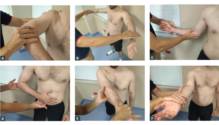
Figure 1. Special tests for shoulder pathology.
A. Assessing for subacromial pain and minor rotator cuff disease with the Hawkins–Kennedy test. The patient’s upper limb is positioned as pictured, and the clinician passively internally rotates the shoulder. If this recreates the patient’s pain, the test is positive. B. Assessing the supraspinatus using Jobe’s (empty can) test. The patient’s upper limbs are positioned as pictured, and the clinician applies downward force, which the patient attempts to resist. Pain or weakness implies a positive test. C. Assessing the infraspinatus. The patient’s upper limb is positioned as pictured, and the patient externally rotates their shoulder against resistance provided by the clinician. Pain or weakness implies a positive test. D. Assessing the subscapularis using the belly press test. The patient’s upper limb is positioned as pictured, and they attempt to press their hand against their abdomen, without moving their elbow. Pain or weakness implies a positive test. Note that if the patient simultaneously extends their shoulder, they are activating their posterior deltoid rather than the subscapularis. E. Assessing for acromioclavicular joint (ACJ) arthropathy using O’Brien’s test (Position 1). The patient’s upper limb is positioned as pictured, and the clinician applies downward force, which the patient attempts to resist. Pain localised to the ACJ that does not change when the forearm is supinated implies a positive test for ACJ arthropathy. Pain that is relieved by supinating the forearm (Position 2), implies a positive test for bicipital–labral pathology. F. Assessing for bicipital–labral pathology using Speed’s test. The patient’s upper limb is positioned as pictured, and the clinician applies downward force, which the patient attempts to resist. Anterior shoulder pain implies a positive test.
Investigations
Plain radiographs
Plain radiographs are recommended for all patients presenting with chronic shoulder pain.12 These are diagnostic in degenerative conditions, including glenohumeral and acromioclavicular joint (ACJ) arthritis, and advanced imaging is not required. X-ray will identify occult fractures and features pathognomonic of past dislocation. A frozen shoulder will have normal X-ray findings.
Ultrasound
Ultrasound for rotator cuff pathology has equivalent sensitivity to magnetic resonance imaging (MRI).13 Asymptomatic rotator cuff tears on ultrasound are common.8,13–18 Results should always be interpreted in conjunction with X-rays and the clinical examination. In older patients, ultrasound can be deferred until after a failed course of conservative treatment.
Magnetic resonance imaging
MRI is indicated in patients with possible massive rotator cuff tears, avascular necrosis, long head of biceps pathology, labral pathology or recurrent dislocation. It might be beneficial in patients with inconsistent examination findings, or mixed pathology. In patients with established arthritis, MRI is unlikely to aid management.17 MRI is highly sensitive and should always be interpreted in the context of the patient’s presentation. Labral tears without clinical evidence of instability are rarely the primary source of symptoms. Counselling patients prior to MRI regarding unimportant findings helps them interpret their own reports.
Blood tests
If clinical assessment raises suspicion for an inflammatory (inflammatory arthropathy or polymyalgia rheumatica), infective or neoplastic process, appropriate blood tests should be ordered. When frozen shoulder is diagnosed, the patient should be screened for type 1 diabetes, thyroid dysfunction and dyslipidaemia due to the strong association between these comorbidities.10
Non-surgical treatment
Although red flag diagnoses or those of diagnostic uncertainty might require more urgent referral, the vast majority of patients with chronic mechanical/nociceptive shoulder pain can be managed with the assistance of physical therapies and activity modification (Table 3).
| Table 3. Suggested management pathways by condition |
| Pathology |
Suggested management pathway |
| Post-traumatic instability |
First-line management
- X-ray
- Prompt orthopaedic referral
|
| Atraumatic instability |
First-line management
- X-ray
- Lifestyle modification
- Physiotherapy
Management of persistent disability at six months
|
| Long head bicep and labral pathology |
First-line management
- X-ray
- Lifestyle modification
- Simple analgesia
- Corticosteroid injection (subacromial or ACJ)
- Physiotherapy
Management of persistent disability at three to six months
- Ultrasound (or MRI if diagnostic uncertainty, or suspected long head biceps/labral pathology or massive cuff tear)
- Orthopaedic referral
(Consider earlier investigation and referral in younger patients with athletic or physically demanding occupations) |
| Subacromial pain and minor rotator cuff disease |
| Rotator cuff arthropathy and major rotator cuff disease |
| ACJ arthropathy |
| Frozen shoulder contracture syndrome |
First-line management
- X-ray
- Lifestyle modification
- Simple analgesia
- Corticosteroid injection (subacromial or intra-articular)
- Physiotherapy
Management of persistent disability at six months
|
| Glenohumeral osteoarthritis |
| ACJ, acromioclavicular joint; MRI, magnetic resonance imaging. |
Education
Patient expectations are a strong predictor of clinical outcome in both surgical and non-surgical treatment of chronic shoulder pain.19–22 Careful communication is important to avoid over-pathologising age-appropriate pathology. Patients should be counselled that satisfactory improvement is likely over a period of months and that surgery is only considered when simple measures have failed.
Lifestyle modification
Advice should be provided about relative rest, positions of comfort and modification of occupational, recreational and personal activities of daily living.
Sleep disturbance is common in patients with chronic shoulder pain. Sleeping propped up on pillows, in a recliner with a pillow underneath the elbow or lying with the upper limb elevated are positions that might provide comfort.23
For women, switching to a properly fitted bra that fastens anteriorly and has wide straps and cross-back support might reduce symptoms in the acute setting.
Asking the patient to keep a symptom diary can assist with identifying activities that are provoking symptoms. Tasks requiring prolonged or repetitive shoulder elevation are often implicated. Devising pragmatic strategies to reduce the total amount of time spent with the shoulder elevated at home or in the workplace might provide relief and prevent symptom recurrence.
Physiotherapy and exercises
There is strong evidence for physiotherapist-led treatment in subacromial pain, rotator cuff disease and frozen shoulder, with successful outcomes in 65–80% of cases.24–28 Physiotherapy focuses on graded strengthening and neuromuscular retraining, and is tailored to an individual’s specific impairments. Even in the setting of severe pain, almost all patients will benefit from supported range-of-motion exercises. Pendulum exercises are often recommended but, in the setting of kinesiophobia, tabletop stretches might be better tolerated. When exercise cannot be tolerated, there is some evidence supporting strapping for short-term pain relief in subacromial pain.29 Where there is limited access to physiotherapy, any exercise (even walking for cardiovascular fitness) can yield positive outcomes for pain and quality of life; however, upper limb resistance exercises performed at high volumes yield the largest improvements in pain and disability.30 For large or massive rotator cuff tears, the goal is to strengthen the deltoid and any intact portions of the cuff. Patients are taught to compensate for loss of cuff function by using ‘short-lever’ strategies to achieve overhead positions. For frozen shoulder, exercises are focused on passive and active stretching of the glenohumeral capsule and rotator cuff and are most effective in the first three months, before true contracture has formed.
Psychosocial support
Catastrophising about symptoms or imaging findings, fear-avoidance behaviours and low self-efficacy are common psychological features that can influence a patient’s outcomes.31 Furthermore, patients with comorbid depression, anxiety or post-traumatic stress are likely to experience poorer musculoskeletal outcomes. It is important to ask patients what their understanding of their condition is and what their coping strategies are. In most cases, unhelpful beliefs and behaviours can be addressed at the point of care with reassurance and education as outlined above. Patients with features of more complex pain psychopathology might need further medical and/or psychological support.2 The use of multimedia, such as the open-access video, Understanding pain in less than 5 minutes,32 can be an efficient way to convey the role of multidisciplinary treatment.
Analgesia
Similar to managing chronic pain in other musculoskeletal conditions, a stepwise approach to analgesia is recommended.33 Regular paracetamol might be beneficial, and a short course of anti-inflammatories should be considered, particularly where the patient is experiencing sleep disturbance. In patients with complex pain needs, referral to a specialist pain service might be required.
Injections
Local anaesthetic and cortisone injections play a diagnostic and therapeutic role in chronic shoulder pathology. Injections can target the subacromial space, glenohumeral joint or ACJ, as well as extrinsic sites, including cervical nerve roots. The patient should complete a pain diary after injection. Even if pain relief is temporary, this provides diagnostic assurance, particularly in the setting of multiple pathologies.
Corticosteroid injections might be trialled for subacromial pain, ACJ arthropathy and arthritis if simple analgesia is ineffective in controlling symptoms.34 In frozen shoulder, there is strong evidence for better outcomes following early corticosteroid injection.24 The frequency might be debated, but one to two injections, spaced six weeks apart is optimal, with unlikely additional benefit to repeat procedures beyond this. The role of biologics, including platelet-rich plasma, is hotly debated but currently not Medicare funded, indicating a lack of reliable evidence on health technology assessment.
Surgical treatment
Surgery might be required for chronic conditions that have failed three to six months of non-surgical treatment. Prior to referral, it is important to inform patients that although surgery might improve pain, it does not always result in complete recovery of premorbid function. Recovery from surgery requires weeks of immobilisation and time off work and driving, followed by several months of rehabilitation before return to pre-injury activity levels.
Although outside of the scope of this article, younger patients with acute rotator cuff tears or traumatic instability should be promptly referred for consideration of repair or stabilisation.27,35
Surgery is an effective treatment for subacromial pain, rotator cuff disease and bicipital–labral pathology. Due to comparable outcomes with non-operative treatment, these surgeries are best considered for patients who have exhausted other treatment options. Large and massive rotator cuff tears, particularly in older patients, are often not viable for repair.
Manipulation under anaesthesia and arthroscopic capsular release are equally effective as physiotherapy for frozen shoulder contracture syndrome.28 Referral for these procedures should only be considered after 6–12 months of non-operative treatment due to high rates of recurrence and the risk of postoperative complications.
Shoulder replacement for advanced glenohumeral arthritis, cuff arthropathy and surgical neck of the humerus fractures are successful operations for older patients.36
Conclusion
Chronic shoulder pain is a common presenting complaint in general practice. Diagnosis requires a thorough history and clinical examination, coupled with appropriate investigations. Most presentations can be successfully managed in the general practice setting with a multidisciplinary approach.
Key points
- Chronic shoulder pain is a common presenting complaint.
- Accurate diagnosis is largely informed by careful history and examination, in conjunction with plain radiographs.
- It is important to interpret imaging in the context of the patient’s history and examination findings.
- Many chronic shoulder conditions can be successfully treated in the general practice setting with a multidisciplinary approach.
- Referral to an orthopaedic surgeon is recommended for certain conditions and for patients who have failed non-surgical management.