Case
A woman, aged 20 years, presented with a seven-year history of progressive asymptomatic hair loss that developed initially on her eyebrows and then progressed to also involve her scalp. There was no relevant past medical or family history and no regular medications. Multiple previous dermatologists had diagnosed either alopecia areata or female pattern hair loss, but the pattern of the hair loss was inconsistent with either diagnosis. The areas of hair loss changed slowly over weeks to months, with some areas extending and others improving. Clinical examination showed multiple broken hairs of variable length with no actual baldness. On trichoscopic examination, there was no inflammation, hair miniaturisation or exclamation mark hairs (Figure 1).
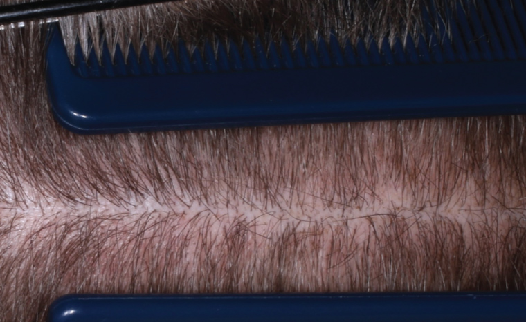
Figure 1. The patient’s hair at presentation.
Question 1
What are four possible differential diagnoses?
Answer 1
Four possible differential diagnoses to consider are trichotillomania, tinea capitis, alopecia areata and androgenetic alopecia. Table 1 outlines the differential diagnoses and clinical reasoning for exclusion for each.
| Table 1. Differential diagnoses with clinical reasoning for exclusion |
| Differential diagnosis |
Clinical reasoning for exclusion in this case |
| Tinea capitis |
- The prolonged duration of symptoms over several years is inconsistent with tinea infection
- The absence of pruritus, scales and corkscrew hairs makes this diagnosis unlikely
|
| Alopecia areata |
- Unlikely because there was no actual baldness in this patient
- In addition, no exclamation mark hairs, which are characteristic of alopecia areata, were seen on trichoscopy
|
| AGA |
- Trichotillomania can mimic AGA, also known as female pattern hair loss, due to their clinical and trichoscopic similarities
- AGA was considered unlikely in this case due to the inconsistent distribution and lack of hair miniaturisation
- Eyebrow involvement is also uncommon in AGA
|
| AGA, androgenetic alopecia. |
Case continued
On further questioning, the patient reported fiddling with her hair, and the patient’s mother commented that she regularly observed her daughter touching and twirling her hair and constantly told her daughter not to fiddle with her hair.
With this new information, the patient was asked to pluck a single hair from her scalp, which she performed expertly. This expertise had been acquired through years of practice. Features visible on close inspection of her scalp that are consistent with a diagnosis of trichotillomania are shown in Figure 2.
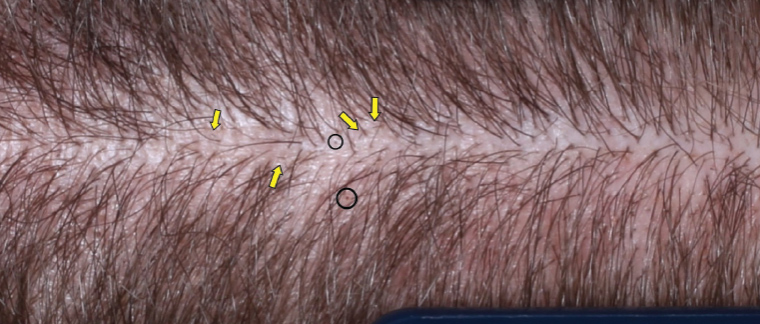
Figure 2. Close-up clinical photograph of the patient’s scalp. Yellow arrows indicate broken hairs of varying lengths. The black dots within the black circles are broken hairs at the level of the scalp.
Question 2
What are the clinical features of trichotillomania on examination?
Question 3
What are the trichoscopic features of trichotillomania?
Question 4
What is a trichobezoar?
Question 5
What is the management of trichotillomania?
Answer 2
The features of trichotillomania on clinical examination include:
- patchy hair loss with angular borders
- hair of varying lengths, with broken brush tip ends seen on close inspection and sometimes felt
- a negative hair pull test1 (see below)
- absence of scalp inflammation
- a positive hair presentation test (see below).
The hair pull test is performed by gently pulling on a bundle of approximately 50–60 hairs; the test is positive when three or more hairs are easily plucked out. A positive hair pull test is indicative of disorders with increased shedding, such as telogen effluvium.2 Trichotillomania is not associated with increased shedding, and therefore the hair pull test is negative in patients with this condition.
The hair presentation test is positive when a patient expertly plucks a single hair and presents it to the doctor.
Answer 3
Trichoscopy, which involves using a dermatoscope to examine the hair and scalp, is an important diagnostic tool. The trichoscopic findings listed in Table 2 are found in trichotillomania, with certain features more sensitive and specific than others. Hairs broken at different levels are seen in nearly all cases; however, this is not a specific finding. Broken hairs are commonly seen in alopecia areata and tinea capitis.3 Trichotilopsis, or ‘split ends’, is the most frequently seen relatively specific finding. Hook hairs and coiled hairs have almost 100% specificity for trichotillomania, although low sensitivity.4
| Table 2. Trichoscopic features of trichotillomania in order of specificity3,4 |
| Trichoscopic features |
Description |
Differentials |
| Hook hairs |
Partially coiled hairs, hook hairs are the most specific trichoscopic feature of trichotillomania |
- Not observed in other condition: 100% specificity for trichotillomania
|
| Coiled hair |
Irregular coil-shaped hairs resulting from pulling force |
- Highly specific to trichotillomania (99.6%)
- Also observed in traction alopecia
|
| V hair |
Two or more hairs broken at the same length emerging from one follicle |
- Characteristic of trichotillomania, with an estimated specificity of 99%
|
| Hair powder |
Almost entirely damaged hair shaft, with only sprinkled hair residue remaining |
- Traction alopecia
- Alopecia areata
|
| Trichotilopsis |
Split ends commonly observed in very short hairs |
- Traction alopecia
- Alopecia areata
- Tinea capitis
- Primary cicatricial alopecia
|
| Flame hairs |
Thin, wavy-shaped hair residue |
- Acute chemo- and radiotherapy-induced alopecias
- Alopecia areata
- Traction alopecia
- Central centrifugal cicatricial alopecia
|
| Broken hairs at different levels |
Numerous hairs broken at different levels
This is seen in nearly all cases of trichotillomania |
- Traction alopecia
- Alopecia areata
- Tinea capitis
|
| Black dots |
Broken hair at the level of the scalp |
- Alopecia areata
- Chemotherapy-induced alopecia
- Dissecting cellulitis
- Tinea capitis
- Traction alopecia
|
Answer 4
Approximately 10–20% of patients with trichotillomania ingest their hair after plucking it, a behaviour known as trichophagia. This can lead to the accumulated hair forming a mass in the stomach known as trichobezoar. A potentially rare but life-threatening complication of trichotillomania, known as the ‘Rapunzel syndrome’, occurs when the trichobezoar extends into the intestines, causing an intestinal obstruction.5
Answer 5
Trichotillomania is included in the disorder class of obsessive–compulsive and related disorders in the Diagnostic and statistical manual of mental disorders, fifth edition (DSM-5).6 The onset of trichotillomania generally begins in late childhood or early adolescence and is often associated with stress, anxiety and depression. The management of trichotillomania involves psychotherapies including cognitive behavioural therapy, habit reversal therapy, dialectical behaviour therapy and exposure therapy.5,7 If stress at home is the trigger for trichotillomania, parents can consider making changes to the child’s environment to reduce stress. In cases where pharmacotherapy is warranted, medications such as tricyclic antidepressants, selective serotonin uptake inhibitors, olanzapine and N-acetylcysteine might be considered.7 Minoxidil can also be used to facilitate hair regrowth. A consultation with a dermatologist or trichologist might be helpful for advice relating to leave-on conditioners and other camouflage aids.
Key points
- The hair presentation test is a simple and effective tool to assist clinicians with making the diagnosis of trichotillomania.
- The management of trichotillomania involves psychotherapy and pharmacotherapy.
- Assessing whether the patient ingests plucked hairs is important to prevent gastrointestinal obstruction.