Foot pain from exercise is frustratingly common and often a reason given by patients for cessation of an exercise program despite improvement in strength, bone health, mood and overall quality of life in people who are active. Foot pain is present in up to 36% of adults and is more common in women, patients aged >55 years and patients who are obese.1 Prompt diagnosis and treatment, particularly focusing on offloading, lifestyle modifications and footwear, using a multidisciplinary team can prevent the development of chronic musculoskeletal pain and expedite recovery and return to activity.
Aim
This paper discusses three common presentations, including diagnosis and management of foot pain following exercise: stress fractures, plantar fasciitis and mid-foot arthritis.
Stress fractures
Stress fractures to the foot, ankle and distal tibia are a common presentation for orthopaedic surgeons and sports and exercise physicians. However, these injuries can also occur in sedentary patients who change their exercise habits or diet. Examples of dietary changes could include deliberate calorie restriction in an attempt to lose weight or systemic disease such as coeliac disease resulting in malabsorption and calorie deficit. Bone turnover is a delicate balance between the accumulation of bone damage and repair, and deviation from regular routine can result in a bony stress response and injury.
Stress fractures in the foot can present as insidious onset of foot pain without a specific history of trauma. A common phenomenon during the COVID-19 pandemic was an increase in stress fractures of the foot and ankle during city-wide lockdowns.2 This was explained by a number of lifestyle changes commonly adopted during the COVID-19 pandemic, including increased uptake in running3 and decreased sunlight exposure and vitamin D deficiency.4 Patients with a stress fracture often complain of night pain, with some patients describing the pain from a stress fracture as ‘a toothache in the foot’. The clinician should take a focused history assessing changes in exercise type and volume. For females of child-bearing age, a menstrual history needs to be obtained because irregular or absent periods can be an indication of increased physiological stress and subsequent risk of stress fracture development.5
Patients will present with point tenderness at the site of the stress fracture. Dorsal foot tenderness directly over the metatarsal shaft is suggestive of a metatarsal shaft stress fracture, with the second metatarsal shaft most commonly involved.6 Navicular stress fractures will often present with point tenderness over the navicular tuberosity on the medial border of the foot, and at the dorsum of the foot distal to the ankle. Calcaneal stress fractures will present with heel pain or pain at the level of the Achilles tendon insertion.
Initial X-rays are often normal and further investigation is warranted if there is clinical suspicion of a stress fracture. Magnetic resonance imaging (MRI) is the gold-standard, least-invasive modality for stress fracture investigation,7 but is not always available, particularly in rural or regional areas. Single photon emission computed tomography (SPECT) is an alternative investigation, particularly in the setting of multiple sources of pain suggesting the potential for two or more stress fractures; alternatively, SPECT might be useful in the diagnosis of systemic disease such as bone marrow oedema syndrome.8
Treatment of stress fractures of the foot or ankle involves treating the injured bone as well as managing the patient systemically. For stress fractures involving the navicular, anterior tibial cortex, fifth metatarsal shaft, sesamoid bones or medial malleolus (Box 1), immediate immobilisation in a controlled ankle motion (CAM) boot or below-knee cast and advice to be non-weight bearing, followed by referral to an orthopaedic surgeon with subspeciality training in the foot and ankle or a sport and exercise physician is appropriate due to the propensity for these injuries to these specific bones to progress to non-union.9 However, most patients with a stress fracture to the remaining bones in the foot or ankle, commonly the metatarsal, will be able to weight bear in a CAM boot on diagnosis.
| Box 1. High-risk stress fractures requiring urgent referral |
- Navicular
- Anterior tibial cortex
- Fifth metatarsal shaft
- Sesamoid bones
- Medial malleolus
|
Treatment also involves addressing the cause of the stress fracture and managing potential systemic pathologies to prevent recurrence. Involvement of a sport and exercise physician might be helpful in managing athletes with stress fractures to assess for relative energy deficiency (RED) syndrome,10 where caloric intake is insufficient for the duration and intensity of training undertaken. RED syndrome can also be associated with menstrual cycle irregularities and poor bone health in older age,11 which reinforces the importance of systemic management for these patients. Assessment of nutritional deficiencies, including eating disorders, food group restriction (eg lactose intolerance) and alcohol intake, should be undertaken.
Initial non-operative management of stress fractures includes immediate cessation of high-impact activity, offloading (usually with a CAM boot) and analgesia. Most stress fractures of the foot and ankle can be successfully offloaded by fully weight bearing in a CAM boot, with the exception of the navicular bone. Referral to an experienced podiatrist or physiotherapist might help address biomechanical deficiencies and formulate a return to a sport or exercise program. A referral to a sport and exercise physician might also be beneficial for elite athletes, or patients undertaking high levels of regular activity.
Stress fractures can fail to heal in up to 10% of lower limb stress fractures.12 In this instance, surgical referral might be warranted. The diagnosis of non-union or delayed union is normally made after six months of non-operative management, and a computed tomography (CT) scan will demonstrate whether cortical bridging is present (Figure 1) and, if no bony bridging is visible, this suggests non-union or delayed union of the fracture. Surgical treatment involves open reduction, internal fixation and bone grafting to the injured tarsal bone (Figure 2) followed by a period of non-weight bearing and rehabilitation, which can be up to six months.
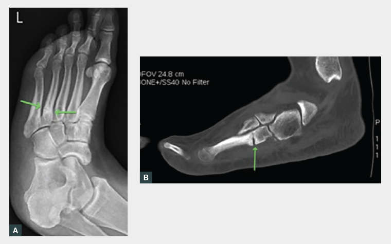
Figure 1. A. A preoperative X-ray of a non-union of a stress fracture to the fourth metatarsal (arrows) in a woman, aged 35 years, who commenced a running program with no prior history of high-impact exercise. B. A preoperative computed tomography scan of the non-union of stress fracture to the fourth metatarsal (arrow) in a woman, aged 35 years, who commenced a running program with no prior history of high impact exercise.
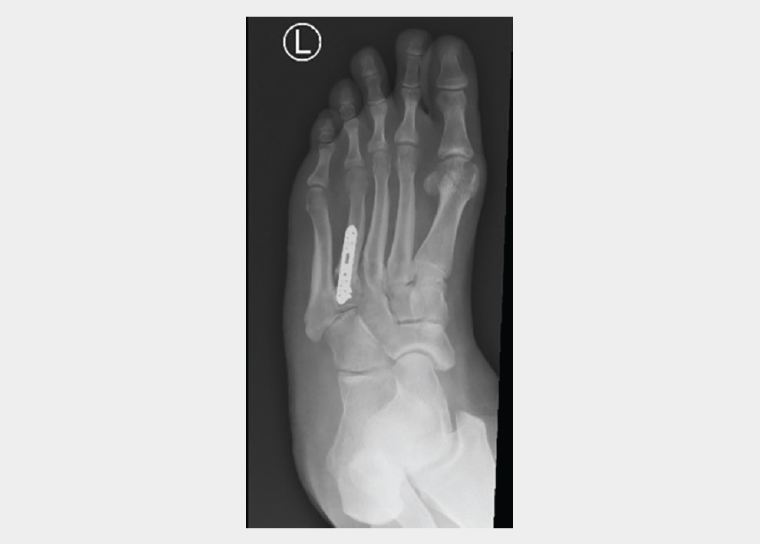
Figure 2. A postoperative X-ray of a successful union of the stress fracture shown in Figure 1, following open reduction, internal fixation and local bone grafting of the fourth metatarsal.
Plantar fasciitis
Plantar fasciitis is a common cause of foot pain following a change in exercise habits. It is caused by chronic inflammation of the plantar fascia aponeurosis on its insertion into the calcaneus and commonly presents with pain in the medial heel. Impact to the plantar fascia can occur with dysfunctional biomechanics, changes in walking surfaces or footwear or changes in exercise habits and walking surfaces. Chronic inflammatory changes and microtears in the plantar fascia can result in chronic fasciopathy and thickening of the plantar fascia.13 The peak age of presentation is between 40 and 60 years and the condition affects men and women equally.14 It is estimated that up to 10% of people will develop plantar fasciitis over their lifetime.15
Risk factors for the development of plantar fasciitis include running athletes, tight gastrocnemius or Achilles tendon contracture and obesity.16 History should focus on activity levels, changes in exercise habits or footwear and change in employment or lifestyle resulting in prolonged standing or weight gain. Patients commonly describe pain first thing in the morning or when arising from a seated position that improves over the course of the day.
Examination demonstrates tenderness to palpation over the medial heel and occasionally mild swelling. Tightness of the Achilles tendon also needs to be assessed and the Achilles tendon palpated at its mid-portion and insertion to assess for potential Achilles tendinopathy. A careful neurovascular examination should be made, in particular assessing the tarsal tunnel and posterior tibial nerve due to the rare pathology of posterior tibial nerve entrapment causing posteromedial heel pain.
The diagnosis of plantar fasciitis might be supported with an ultrasound to demonstrate thickening or partial tears of the plantar fascia.17 Although not a first-line investigation, an MRI can be useful in patients with neurological symptoms, refractory symptoms or for patients with concomitant pathology such as achilles tendinopathy. In particular, an MRI scan will demonstrate a tarsal tunnel ganglion that, if compressing the posterior tibial nerve, can present in a similar manner to plantar fascitis. Very rarely, a calcaneal stress fracture can present as heel pain and a calcaneal stress fracture would also be diagnosed via MRI. The use of X-ray and the presence of a ‘heel spur’ in the diagnosis of plantar fasciitis is controversial. A heel spur is evident arising from the plantar aspect of the calcaneus on a lateral X-ray in only 50% of patients with plantar fasciitis,16 so interpretation of the X-ray needs to be made with caution to avoid misdiagnosis.
Treatment of plantar fasciitis is multidisciplinary and can be frustrating for both the treating clinician and patient. The average recovery period has been reported to be as long as 12 months in some studies, but patients should be reassured that 90% of patients will improve.16 Modifying activities to avoid running and prolonged standing and weight management are important. Simple analgesia, such as paracetamol and an anti-inflammatory, can provide some relief. Engaging a physiotherapist or podiatrist might be useful to trial low dye taping, Achilles tendon and calf stretching and footwear changes. In addition, allied health practitioners can assess biomechanics, load management and exercise rehabilitation to facilitate a return to exercise and prevention of recurrence of the condition. There might be a limited role for extracorporeal shock wave therapy. A recent randomised controlled trial reported improvement in heel pain in 50–65% of patients undergoing shock wave treatment for insertional Achilles tendinopathy,18 although the results of that paper needs to be extrapolated to heel pain from plantar fasciitis with caution. Injection of a local anaesthetic and steroid, as well as dry needling, have also demonstrated some improvement in plantar fasciitis symptoms.19 With so many treatments for plantar fasciitis, with varying levels of effectiveness, cost and availability, it is understandable that this is a frustrating condition for patients and clinicians.
There might be a limited role for surgical release of the plantar fascia, usually performed endoscopically, in severe, refractory cases. However, surgical intervention is not always reliable, with a recent systematic review reporting ‘weak evidence to support that endoscopic plantar fascia release was safe and effective for the treatment of chronic plantar fasciitis’.20 A referral to an orthopaedic surgeon for surgical management of plantar fasciitis should be the final treatment modality after all non-operative measures have been exhausted.
Arthritis in the foot
Arthritis of any of the 33 joints in the foot is common.21 A recent study assessing the management of foot and ankle osteoarthritis by general practitioners (GPs) in Australia suggested this condition accounts for an estimated 152,000 general practice encounters nationally, mostly in patients aged 65–74 years.22 Patients might not report pain at the time of activity, but commonly report either pain in the evening or following day that might limit walking or a limp. There might be a history of trauma or injury to the foot or ankle.
Examination can demonstrate stiffness of the arthritic joints and often pain with attempted mobilisation. Patients will usually have an antalgic gait, particularly when arising from a seated position. The arthritic joints are usually tender to direct palpation and patients will often complain of a dull ache even after pressure has been removed from the irritable joint. The presence of osteophytes will present as a bony prominence on the dorsum of the foot in tarsometatarsal joint arthritis or the hallux in first metatarsophalangeal joint arthritis. These can be very irritating and rub on shoes even though the underlying arthritis might not be painful. Arthritis to the foot usually presents in the later decades, so peripheral examination, including assessing for pulses and sensation, is important due to the risk of underlying peripheral vascular disease and/or diabetes.
A weight-bearing X-ray is the mainstay of diagnosis in arthritis of the foot and ankle. The majority of moderate to severe arthritic conditions will be easily diagnosed via plain radiographs and further imaging is not required. Mild, moderate and severe arthritis can be demonstrated via CT scan or, if inflammatory arthritis is suspected, an MRI will demonstrate synovitis, perichondral erosions and cyst formation that can indicate inflammatory arthritis such as rheumatoid arthritis rather than osteoarthritis. An MRI scan will also demonstrate concomitant pathologies such as tendinopathy, ganglion cysts and neuromas.
Treatment of arthritis of the foot involves activity modification, weight loss and footwear changes. Prescription rates by GPs exceeded non-pharmacological interventions such as counselling, advice or education or allied health referral.22 Allied health practitioners such as podiatrists and physiotherapists are experienced in the management of foot and ankle arthritis and an exercise physiologist might help modify activity to facilitate weight loss in the setting of arthritis. Offloading the arthritic foot with a rocker-bottom shoe might be helpful, particularly in patients with arthritis of the mid-foot and/or the forefoot, and these shoes have become mainstream and accessible, and have even demonstrated to help running performance.23 The role of steroid injections in arthritic joints of the foot and ankle is well established and they have been shown to decrease pain and delay surgical intervention.24 Ultrasound-guided injections are recommended due to the small joint space and complex anatomy of the foot. Injections can be useful for both diagnostic and therapeutic purposes, but should be reserved for patients who have a limited response to less invasive treatment methods. The frequency of steroid injections is dependent on the initial effectiveness of the injection, as well as patient factors, such as needle phobia.
Involvement of an orthopaedic surgeon with speciality training in foot and ankle surgery is appropriate when non-operative measures have failed. In the absence of pain but irritability from dorsal osteophytes, a simple bumpectomy might be appropriate to remove the offending bumps and improve footwear options for patients. However, this does have the risk of recurrence of development of pain from arthritis in the future. Alternatively, the majority of arthritic joints in the foot can be treated with an arthrodesis (fusion). The recovery from this procedure varies according to the exact joint that is fused but will usually involve a two-week period of non-weight bearing followed by a further four-week period of non-weight bearing or partial weight bearing in a CAM boot, cast or postoperative shoe. Patients generally return to regular walking three months after their procedure. In the author’s experience, patients are often concerned about losing range of motion of the fused joint(s), but clinical studies demonstrate a near-normal gait following fusion of the first metatarsophalangeal joint, with less pain and increased walking distance.25 An arthrodesis to the mid-foot is still a reliable procedure, although it has a higher complication rate compared with forefoot procedures.26
Conclusion
These three common causes of exercise-induced foot pain are often best treated via a multidisciplinary team following accurate diagnosis from a patient’s GP. Footwear changes, lifestyle modifications and allied health involvement are appropriate initial management for the majority of patients presenting with foot pain after exercise. Referral to an orthopaedic surgeon is appropriate should initial non-operative management not be successful or if the GP has any concerns about their patient’s progress.