Case
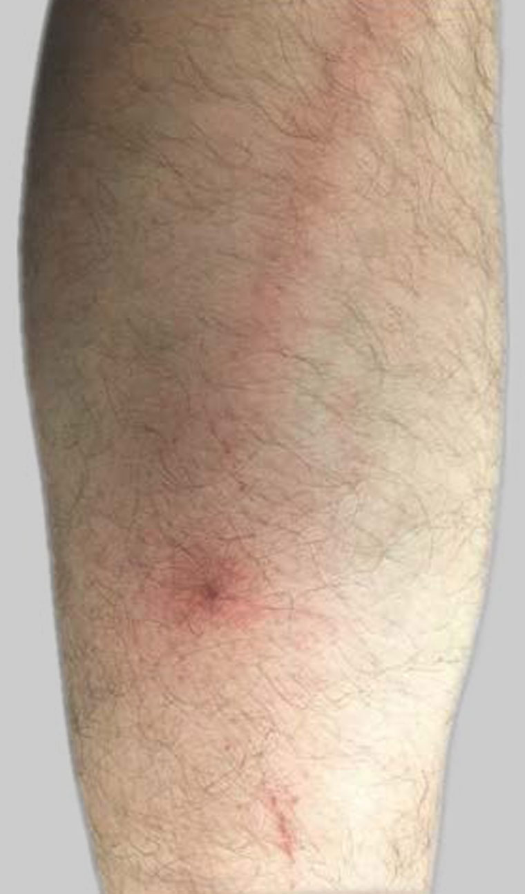
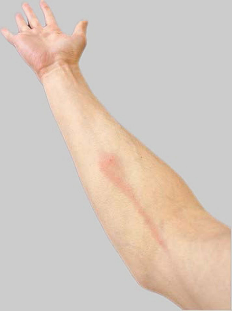
A man, aged 31 years, wakes to find a pruritic erythematous papule on the anterior aspect of his right forearm, approximately 5 mm in diameter, with a central haemorrhagic punctum. The previous day he had been gardening in his metropolitan Australian home when he felt something crawling on the same right forearm, which was promptly squashed and hence subsequently unidentifiable. Over the following 24 hours, despite refraining from scratching the papule, he developed a well-demarcated erythematous linear streak originating from the papule and extending proximally to the cubital fossa (Figure 1). There was no associated pain, tenderness on palpation, induration, palpable cord-like thickening or palpable lymphadenopathy. The patient was systematically well and afebrile. He had no past medical history and no regular medications. However, five years prior, he recalled a history of a similarly pruritic papule followed by streak-like erythema on his lower leg, again following gardening (Figure 2).

Figure 1. A well-demarcated, linear erythematous streak originating from a papule with a central, crusted punctum on the anterior forearm of the patient 24 hours after the papule was first noted.

Figure 2. Previous leg lesion (five years prior) demonstrating a well-demarcated, linear erythematous streak, originating from an erythematous papule with a central crusted punctum, extending proximally along the posterior aspect of the calf.
Question 1
Which types of differential should be considered for this skin lesion?
Question 2
What are the possible aetiologies of superficial lymphangitis?
Question 3
How would you treat bacterial lymphangitis?
Question 4
How can you treat arthropod-associated superficial lymphangitis?
Answer 1
The well-demarcated linear region of erythema raises several possibilities. In particular, superficial lymphangitis and superficial thrombophlebitis are relevant considerations. In this instance, the presumed mode of acquisition, the lack of a palpable vein, the short duration of time between the bite and development of erythema, and the lack of systemic symptoms make arthropod-associated superficial lymphangitis the most likely diagnosis. Phytophotodermatitis may be considered as a differential; however, the distribution of the erythema in this case clearly extends from the punctum and follows the course of the lymphatic vessels, supporting the diagnosis of lymphangitis.
Answer 2
Superficial lymphangitis can be caused by both bacterial and non-bacterial aetiologies. Classical bacterial causes of acute lymphangitis (Table 1) include Streptococcus pyogenes and Staphylococcus aureus.1 Bacterial lymphangitis can be associated with clinical features such as fever and local lymphadenopathy.2 Non-bacterial causes of acute superficial lymphangitis are diverse and less common. These aetiologies include viral (particularly from herpes simplex), fungal (particularly from Sporothrix schenckii) and parasitic infections, as well as arthropod-associated lymphangitis (Table 1).3
The pathophysiology of arthropod-associated superficial lymphangitis remains unknown; however, it is believed to involve a toxic or allergic process induced by the secretions of athropods.4 It is thought that a linear spread of the inflammation occurs due to the contiguous diffusion of the toxin and/or the inflammatory mediators through the lymphatics adjacent to the dermis.4 Previously reported cases describe that it is not uncommon that the offending arthropod cannot be identified, or that a bite itself was not observed.5 However, when identifiable, the implicated arthropods have included mosquitos, spiders and scorpions.6,7
|
Table 1. Comparison of the clinical characteristics and management of arthropod-associated superficial lymphangitis, acute lymphangitis, herpes simplex lymphangitis and sporotrichoid lymphocutaneous infection
|
| |
Arthropod-associated superficial lymphangitis
|
Acute bacterial lymphangitis
|
Herpes simplex lymphangitis
|
Sporotrichoid lymphocutaneous infection
|
|
Typical presentation
|
- Linear erythematous streak originating from an erythematous macule or papule with a central haemorrhagic punctum, extending proximally along the course of a lymphatic vessel3,4
|
- Linear erythematous streak extending proximally along an extremity, following the course of a lymphatic vessel1,2
|
- Vesicular lesions associated with lymphangitic streaking
- May occur secondary to herpetic whitlow3
|
- Nodular lymphangitis involving multiple asymptomatic (or mildly tender) nodules extending in a linear fashion along the infected arm or leg
- Nodules may ulcerate8,9
|
| Aetiology/associations |
- Arthropod bite (non-infectious)3
|
- Bacterial infection with cellulitis, most commonly Streptococcus pyogenes2,7
|
- Viral infection from herpes simplex virus3,10
|
- Fungal infection from Sporothrix schenckii8,9
|
| Symptoms |
- Usually pruritic and non-tender7
|
- Usually tender or painful2
|
- May be tender
- or painful3,11
|
- Usually tender or painful8,9
|
| Fever or systemic symptoms |
|
|
|
|
| Lymphadenopathy |
|
|
|
|
| Treatment |
- Self-limiting
- Consider symptomatic treatment with topical corticosteroids and/or oral antihistamines if required3
|
- Antibiotic therapy and analgesia1,2
|
- Self-limiting: natural history of 14–21 days10
- Early antiviral therapy can be considered10,11
|
- Oral potassium iodide solution or oral antifungal agents8,9
|
Answer 3
Typically, bacterial lymphangitis requires an antibiotic regimen with empirical streptococcal and staphylococcal coverage, such as flucloxacillin.1,2 Less commonly, lymphangitis may be caused by alternative bacteria, such as Pasteurella multocida, which can be acquired through cat and dog bites.3 Empiric antimicrobial therapy for soft tissue infections associated with cat and dog bites typically includes antibiotics such as amoxicillin–clavulanate. Populations with special considerations may require individualised antibiotic regimens, such as greater Gram-negative bacterial coverage in immunocompromised individuals.
Answer 4
Arthropod-associated lymphangitis has previously been an under-recognised cause of such presentations. In the literature, a number of cases have been described in case series, demonstrating that this diagnosis is a distinct pathological entity.4,7 Furthermore, these cases have been managed successfully without antimicrobial therapy. In these cases, patients were managed successfully with antihistamines and/or topical corticosteroids.7 Such described cases managed without antimicrobials have also included paediatric populations.
In the case of presumed arthropod-associated superficial lymphangitis, clinical monitoring for progression or the development of systemic symptoms is required irrespective of antimicrobial prescription. In a patient for whom the diagnosis was unclear, or who could not be reliably monitored, the decision to prescribe an empirical antimicrobial course may be reasonable. However, in appropriately selected cases, monitoring without antimicrobials could be undertaken following a risk-versus-benefit discussion with the patient, as has been described previously.7 Relevant factors to consider include the patient’s comorbidities, immunosuppression, health literacy and likelihood of adherence to instructions.
Case continued
In this case, it was considered that the diagnosis was most likely arthropod-associated lymphangitis. The inability to identify the original culprit organism limited diagnostic evaluation further. The patient was managed conservatively. Close clinical monitoring was used. Over the following day, the erythema extended approximately a further 5 cm proximally, before gradually resolving over two to three days. No antimicrobials were required. Counselling was undertaken regarding appropriate gardening attire to help minimise the risk of recurrence.
Key points
- Linear erythematous lesions have a differential diagnosis including lymphangitis, superficial thrombophlebitis and allergic contact dermatitis.
- Lymphangitis can be due to bacterial infection, non-bacterial infection and non-infectious aetiologies.
- Although primary bacterial infection or superinfection should always be carefully considered, cases of arthropod-associated lymphangitis can be successfully managed without antimicrobial therapy.