Case
A man aged 60 years presented with an enlarging penile lesion that had been present for six years. He did not have pain, any symptomatology or erectile dysfunction. There was no history of sexually transmissible infections. He was in a monogamous heterosexual relationship of 20 years’ duration. The patient’s medical history was unremarkable, and he took no medications. On examination, a solitary 1 cm × 2 cm scaling keratotic plaque was present on the glans penis. It was indurated but non-tender (Figure 1).

Figure 1. Patient with penile lesion
Question 1
What are the likely diagnoses?
Question 2
What would be your immediate management?
Answer 1
The history and presentation are most consistent with verrucous carcinoma or the more serious penile squamous cell carcinoma. A hyperkeratotic viral wart is also possible.1
Answer 2
The first step is to confirm the nature of this lesion by representative biopsy. Options include a shave, incisional or punch biopsy. This is preferably performed with local infiltration of 1% lignocaine with 1:100,000–1:200,000 adrenaline using as small a dose as possible.2 Adrenaline-containing local anaesthetic is routinely and safely used in penile surgery.3 Suturing typically provides immediate haemostasis for incisional or punch biopsies. Regional lymph nodes should be palpated. Ultrasonography is indicated if squamous cell carcinoma is confirmed.
Case continued
A deep and wide shave biopsy was performed. This was reported as an 18 mm × 8 mm × 6 mm specimen revealing a verrucous carcinoma extending to all margins.
Question 3
What are the risks with penile biopsies?
Question 4
What factors influence the risk for developing male genital neoplasia?
Question 5
What is the significance of the diagnosis of verrucous carcinoma?
Question 6
What treatment options are available for verrucous carcinomas?
Answer 3
Penile skin biopsies are generally safe procedures with minimal complications. Patients should be made aware of the pain associated with injection of anaesthetic at this site and risk of syncope. The patient should be positioned for surgery with this risk in mind. Adrenaline-containing local anaesthetic is safe for local penile injection.3,5
There are risks common to all skin biopsies including bleeding, wound infection, wound dehiscence and permanent scarring. Excessive bleeding is uncommon but more likely to occur in patients taking antiplatelet and anticoagulant medications. Infection is uncommon, but patients with diabetes and those who are immunosuppressed are at increased risk.6 Hypersensitivity to local anaesthetics may occur.7 However, local anaesthetic is generally very safe to use.3,8
Male genital biopsy carries minimal site-specific risks. There is a risk of damage to the urethral meatus with a biopsy, and scarring at this site can result in inability to accurately direct urinary stream. The risk of damage to underlying corpora cavernosum tissue is negligible as it lies deeper than a standard skin biopsy could reach, and such damage has not been reported.9 Post-procedure, there may be prolonged pain or discomfort. Rarely, damage to surrounding nerves can lead to temporary or permanent sensory loss.9
Answer 4
Male genital cancer most commonly affects patients aged 50–59 years. Presence of a foreskin, phimosis, poor hygiene habits, heavy cigarette smoking, human papillomavirus (HPV) infection, psoralen and ultraviolet A (PUVA) phototherapy, immunosuppression and obesity are known risk factors. Scrotal squamous cell carcinoma has been specifically associated with coal tar and chronic mechanical irritation.
4 Neonatal/childhood circumcision and HPV vaccination are associated with a decreased risk.
7
Answer 5
Verrucous carcinoma is a rare variant of well-differentiated squamous cell carcinoma associated with HPV infection. Most commonly occurring on the penis, it has also been reported in the oral cavity, anus and female genitalia.1 It is slow growing and locally aggressive but rarely metastasises. It is considered less dangerous than other squamous cell carcinomas and has a favourable prognosis, and inguinal lymphadenectomy is rarely indicated.1 Computed tomography of the pelvis may be used to evaluate inguinal and pelvic lymphadenopathy in higher-risk malignancies.
Invasion and damage to underlying soft tissue may occur, and occasionally perineural, muscle and even bone invasion. Early diagnosis and prompt management of this lesion is needed.10
Answer 6
Treatment of penile verrucous carcinoma is surgical excision. Most cases should be referred to a urologist or plastic surgeon for surgical management and consideration of further work-up. Consensus is still lacking for the surgical margin required. Typically, a 5 mm margin is used.11 It is a site at which tissue conservation and preservation of form and function is important.
The role of radiotherapy is controversial because of the inability to confirm histological margin control and long-term radiation changes with this modality.12 Radiation therapy should only be employed in cases deemed not amenable to surgery.12
Local surgical resection is the mainstay of treatment. Full or partial penectomy is rarely needed and is reserved for repeated local recurrence or large or deeply invasive lesions. This ensures penile function is preserved where possible.1
Treatments such as topical 5-fluorouracil cream, imiquimod or photodynamic therapy have no role in invasive malignancy.9
Case continued
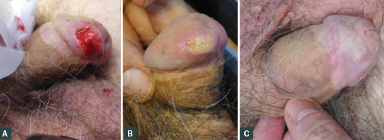
As verrucous carcinoma has an extremely low risk of metastasis, computed tomography of the pelvis was not performed. Further excision revealed no residual lesion. The patient remained recurrence free six years post-operatively. Figure 2 illustrates his management.

Figure 2. Lesion on the glans penis
A. Immediately post-biopsy; B. Two weeks post-biopsy; C. Four years post-treatment
Key points
- Penile biopsies are safe procedures.
- Lignocaine and adrenaline can be safely used for penile biopsies.
- Verrucous carcinoma is a low-risk malignancy amenable to simple excision.
- Local resection is the mainstay of management of invasive malignancy at this site.