Case
A 68-year-old woman requests volumetric modulated arc therapy (VMAT) treatment of her solar damage. Her first basal cell carcinoma was treated at age 37 years. At subsequent three-monthly reviews, numerous basal and squamous cell carcinomas have been surgically treated on her face, trunk and limbs. Cryotherapy and 5-fluorouracil cream have been used for keratoses. She takes no medications, is physically active and remains in part-time work.
Her relative, with ‘field cancerisation of his skin, like me’, recently had VMAT to the face, scalp and limbs with ‘great results’.
Question 1
What is VMAT?
Question 2
What is meant by ‘skin field cancerisation’?
Answer 1
A form of radiation treatment, VMAT delivers external beam radiation treatment more precisely to the target while limiting exposure of normal tissue. It allows more even treatment of rinds of skin regardless of contour while sparing deeper normal tissue and important nearby structures.
Dosage can be uniform throughout the field or with localised ‘boost’ to identified malignancies. Acute and long-term biological effects of radiation on treated tissue remain the same as older techniques (Table 1).1 VMAT is promoted in the medical literature and in the media for field cancerisation.2,3
| Table 1. Possible adverse effects of volumetric modulated arc therapy1 |
| Acute |
Chronic |
| Acute radiation dermatitis (ie erythema, pruritus, dry or moist desquamation) |
Chronic radiation dermatitis/fibrosis |
| Skin ulceration: uncommon, but increased risk with very large fields and high doses |
Poikiloderma (ie atrophy, dyspigmentation, telangiectasias) |
| Oedema and pain, especially when treating most of the circumference of a limb |
Lymphoedema (when treating most of the circumference of a limb) |
| |
Alopecia, reduced sweating |
| Radionecrosis (rare) |
| Induction of secondary malignancy (rare) |
| Impaired range of motion when treating over joints |
Answer 2
‘Skin field cancerisation’ refers to an area of solar damage at increased risk of skin malignancy. The diagnostic criteria are currently poorly defined (Table 2).4–6 Not all definitions require past or present skin malignancy, or visible solar keratosis. Efforts are underway to produce a reproducible grading system for field cancerisation.
| Table 2. Current definitions of skin field cancerisation from the literature |
| Reference |
Definition |
| Willenbrink et al4 |
Multifocal clinical atypia characterized by actinic keratoses or squamous cell carcinomas in situ with or without invasive disease, occurring in a field exposed to chronic ultra-violet radiation |
| Figueras Nart et al5 |
Field cancerization is clinically defined as the anatomical area with or adjacent to actinic keratoses and visibly sun damaged skin identified by at least two of the following signs: telangiectasia, atrophy, pigmentation disorders and sand-paper [sand-paper-like texture]. It is unclear if a visible actinic keratosis lesion is needed for field cancerization |
| Christensen et al6 |
A practical working definition of field cancerization requires three features: a defined region of skin, multiple actinic keratoses within that region and at least one prior squamous cell carcinoma |
Case continued
Examination reveals severe solar damage over skin exposed when wearing a bikini. There are numerous treatment scars, solar keratoses and two basal cell carcinomas (Figure 1).
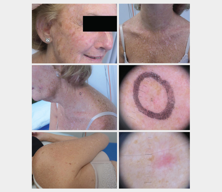
Figure 1. Photomontage of a woman with significant solar damage with numerous solar keratoses and two small basal cell carcinomas.
Question 3
What factors could have precipitated the patient’s interest in VMAT?
Answer 3
Severe solar damage is a chronic disease. Management imposes significant physical, emotional, time and financial burdens. Three decades of active treatment can generate multiple sources of dissatisfaction (Table 3).7,8
| Table 3. Possible sources of dissatisfaction7,8 |
| Frequency of skin examinations and procedures |
| Anxiety over past and/or future skin malignancies |
| Current treatment strategies |
Surgery-related physical discomfort
and/or restriction of activity |
| Solar damage itself and disfigurement of scars from treatment |
| Financial burden from medical costs and time away from the workplace |
Case continued
The patient finds frequent skin checks, followed by periods of relative disability secondary to surgery, problematic. She hopes VMAT will reduce the need for skin checks and surgery.
Question 4
What other management options are available?
Answer 4
Optimal management needs to be individualised. There are a number of interventions that may reduce development of new skin malignancies. Treatment options for skin malignancy are numerous. Selection balances the sometimes conflicting demands of cure, convenience, cost and cosmesis (Table 4).9
| Table 4. Management strategies to reduce the risk of developing new skin malignancies and optimise treatment9 |
| Sun protection measures, including reduction in time spent outdoors |
| Regular surveillance (crucial for the early detection and treatment of new malignancies) |
| Topical field-directed treatments, such as keratolytics and retinoids |
| Topical 5-fluorouracil (with or without calcipotriol); this has been shown to decrease the development of squamous cell carcinoma |
| Other field treatments such as imiquimod, daylight and lamp-based photodynamic therapy, chemical peels and laser resurfacing |
| Oral chemoprophylaxis with vitamin B3 or acitretin |
| Use of lesion-directed non-surgical modalities for suitable individual malignancies, such as 5-fluorouracil, imiquimod, photodynamic therapy and radiation treatment |
| Physical therapy approaches to skin malignancies other than excision, such as cryotherapy or curettage and cautery; these allow same-day treatment of multiple malignancies |
| If the patient finds local anaesthetic surgery confronting, measures such as sedation or even general anaesthesia could be offered |
Case continued
After consultation with the radiation oncologist, the patient is undecided about further management.
Question 5
What would the radiation oncologist have discussed with her?
Answer 5
The patient’s ‘field cancerisation’ has been managed for over three decades. She remains well, employed and active. There are multiple options for management (Table 4). Given the nature of radiation treatment, careful consideration of risks and benefits is crucial. The decision to use VMAT is optimally considered by a multidisciplinary team including plastic surgery, dermatology and radiation oncology. Management needs to be individualised and accompanied by informed consent.
Field radiation is a useful modality in selected patients. The optimal indications for field radiation are not well defined. It is an option where the burden of skin malignancy in terms of frequency, severity and number of lesions is such that usual measures are overwhelmed or create excess morbidity. The patient’s life expectancy needs to be such that the risk of impact from long-term side effects is minimised. Traditionally, radiation treatment of skin lesions has been restricted to patients over 60 years of age to limit the potential for long-term side effects.10
The benefits and risks of ionising radiation for in situ and invasive non-melanoma skin malignancies, such as squamous, basal and Merkel cell carcinomas, are well established. Cure rates for suitable skin malignancies are excellent.11
In small unblinded studies with short follow-up durations, VMAT for field cancerisation has been reported to produce good functional and cosmetic outcomes.10,12 Indications for VMAT include:
- extensive ‘cancerisation’ across large fields or a high tumour load
- a high risk of recurring lesions
- patients who are not surgical candidates
- failure of other therapies.
Sterilisation of actinic keratoses and durable prevention of subsequent skin malignancy is not proven.
Long-term effects develop over many years and are typically irreversible and occasionally progressive. Large VMAT fields have been complicated by treatment-resistant dermatitis, especially if on the lower limbs. Invasive skin malignancy can arise in treatment fields. Induction of malignancy is a risk.13
Multiple cancers arising in treated fields have been seen.13,14 Surgery in irradiated skin is hampered by decreased tissue mobility and risks of wound breakdown.15 Further radiation to a treated field is contraindicated, except in exceptional circumstances, due to the risk of radionecrosis.16 This may compromise the management of patients who develop malignancies with perineural spread or Merkel cell carcinoma.
A common VMAT regimen for field cancerisation requires five treatments per week for five weeks. This can result in considerable out-of-pocket costs. Geographic isolation limits access to VMAT. Evidence-based consensus on the indications for and optimal regimen of VMAT is lacking. Sufficiently powered long-term prospective studies comparing the efficacy and safety of VMAT with current best practice do not exist.
The radiation oncologist would explain these factors to the patient and suggest review within a multidisciplinary clinic to explore all options.
Case continued
The basal cell carcinomas identified were treated with curettage and cautery the day they were found. The patient is uncertain regarding the future management of her field cancerisation. She has been advised to continue with sun protection, skin self-examination and ongoing surveillance.
Key points
- Current definitions of field cancerisation cover a wide range of solar damage.
- VMAT does not have a clearly established role in the management of field cancerisation. Suitably powered, controlled, long-term trials are needed before its routine use can be recommended.
- There are many options for the management of field cancerisation. Careful consideration by a multidisciplinary team is needed.